
脫唇痣
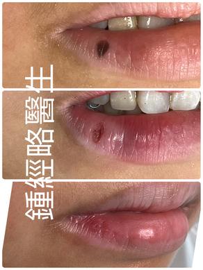
圖1(脫唇痣術前):
脫唇痣 之前:右下唇紅唇緣可見一界限清楚、扁平、深褐色至黑色的斑疹樣病灶,直徑約3–4 mm,色素分佈均勻,表面光滑,無潰瘍、脫屑或邊界不規則等表現。鑒於該病灶位於高可視度的美學區域,且存在因日常摩擦引發刺激或患者心理顧慮之可能,故決定施行手術切除-脫唇痣。
圖2(脫唇痣術後,CO₂雷射切除後):
脫唇痣 之後的傷口:病灶已採用二氧化碳(CO₂)雷射精準汽化切除,形成一處淺表、邊界清晰的凹陷性創面,位於紅唇組織內。創面無癡皮形成,亦無活動性出血,符合CO₂雷射良好的止血特性。隨後使用可吸收縫線,以精細的間斷縫合法進行閉合,確保創緣對合良好、張力最小化。創周可見輕度紅斑,屬術後正常的急性發炎反應。
脫唇痣術後護理
溫和清潔:每天數次以無菌水或溫和的抗菌溶液沖洗。
保持傷口濕潤:使用凡士林或抗生素藥膏保持濕潤;避免乾燥,因為乾燥會導致結痂並延遲癒合。
避免刺激:不要摳痂;避免食用辛辣、酸性或過熱的食物,以免刺激傷口。
口腔衛生:保持良好的口腔衛生以減少細菌量。刷牙時要輕柔,避免造成創傷。
防曬保護:使用護唇防曬或避免直接曝曬,因為紫外線可能導致色素沉著。
監測感染:注意是否有紅腫、膿液或疼痛加劇。如有這些情況,應立即就醫。
圖3(拆線後):
於拆線後即刻拍攝,創面已完全上皮化,僅遺留一條極細微、略呈輕度色素減退的線狀疤痕。在自然光下仔細觀察方可察覺,整體與周圍紅唇組織色澤及輪廓高度融合,幾乎不可見。唇部形態及功能完全保留,未見攣縮、肥厚性疤痕或色素異常等併發症。此結果充分體現CO₂雷射精準切除聯合精細縫合技術在高可視區域(脫唇痣)實現卓越美容效果的能力。
Figure 1 (Preoperative):
A well-circumscribed, flat, dark brown to black macular lesion is visible on the right lower vermilion lip. The lesion measures approximately 3–4 mm in diameter, with uniform pigmentation and smooth surface texture. No signs of ulceration, scaling, or irregular borders are present. Given its location in a highly visible aesthetic zone and potential for trauma or patient concern, surgical removal was indicated.
Figure 2 (Immediate Postoperative – Following CO₂ Laser Excision):
The lesion has been precisely ablated using a carbon dioxide (CO₂) laser, resulting in a shallow, well-demarcated depressed wound bed within the vermilion tissue. Notably, there is no scab formation or active bleeding at the site, consistent with the hemostatic properties of the laser. The defect was closed using fine, interrupted absorbable sutures placed with meticulous technique to ensure optimal edge approximation and minimize tension. Mild surrounding erythema is present, reflecting an expected acute inflammatory response.
Immediate Post-Procedure Care
Gentle Cleaning: Rinse with saline or mild antiseptic solution twice daily.
Keep the wound moist with petroleum jelly or antibiotic ointment: Avoid dryness, which can lead to crusting and delayed healing.
Avoid Irritation: Do not pick at scabs; avoid spicy, acidic, or very hot foods that can sting the wound.
Oral Hygiene: Maintain good oral hygiene to reduce bacterial load, but brush gently to avoid trauma.
Sun Protection: Use lip sunscreen or avoid direct sun exposure, as UV can cause pigmentation changes.
Monitor for Infection: Watch for redness, pus, or increasing pain. Seek medical attention if these occur.
Figure 3 (Post-Suture Removal):
Photographed immediately after suture removal, the wound demonstrates complete epithelialization with minimal residual scarring. A faint, linear, slightly hypopigmented scar is visible upon close inspection, but it is virtually imperceptible under natural lighting and blends seamlessly with adjacent vermilion tissue. Lip contour and function remain fully preserved, with no evidence of contracture, hypertrophy, or pigmentary disturbance. This outcome underscores the precision of the laser excision and the effectiveness of the fine suture technique in achieving an excellent cosmetic result in a high-visibility area.