Discussion: Surgical Challenges and Clinical Considerations in CO2 Laser Ablation of a Cutaneous Upper Lip Nevus
討論:CO2 雷射氣化切除上唇皮膚區域黑色有毛痣的手術挑戰與臨床考量
Clinical Description of the Lesion
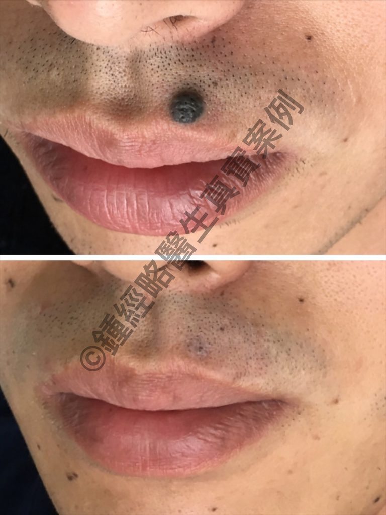
- Location: The lesion sits on the left upper cutaneous lip, positioned superior to the vermilion border and medial to the nasolabial fold.
- Morphology & Type: It presents as a solitary, hyperkeratotic, heavily pigmented exophytic papule/nodule. Additionally, the surface displays a distinct verrucous (wart-like) or cerebriform architecture.
- Color: The body of the lesion exhibits uniform, dark brown to jet-black pigmentation.
- Borders & Shape: It features a sharply circumscribed, well-demarcated circular to oval shape with smooth, regular peripheral margins.
- Size: The lesion measures approximately 5–6 mm in maximal diameter.
- Associated Features: Multiple coarse, dark terminal hairs protrude directly from the lesion surface, confirming a hairy nevus with hypertrichosis.
- Surrounding Tissue: The adjacent skin shows normal perioral facial stubble and mild creases. Crucially, it lacks any signs of surrounding erythema, ulceration, active bleeding, or satellite lesions.
Clinical Impression: The findings strongly suggest a benign intradermal or compound melanocytic nevus (verrucous/pilose type).
這粒墨痣的皮膚科描述:
- 部位(Location): 位於左側上唇皮膚區域,處於唇紅緣(Vermilion border)上方,鼻唇溝內側。
- 型態與類型(Morphology & Type): 單發、過度角化(Hyperkeratotic)且高度色素沉著的外生性丘疹/結節(Exophytic papule/nodule),表面呈疣狀(Verrucous)或腦回狀(Cerebriform)結構。
- 顏色(Color): 呈深棕色至漆黑色,病變本體色素沉著均勻。
- 邊界與形狀(Borders & Shape): 邊界清晰(Well-demarcated)、界限分明,呈圓形至橢圓形,外周邊緣平滑且規則。
- 大小(Size): 最大直徑約 5–6 mm。
- 伴隨特徵/毛囊皮脂腺(Associated Features): 病變表面可見粗硬的深色終毛(Terminal hairs)直接穿出,符合有毛痣/伴多毛症之複合痣或真皮內黑素細胞痣(Compound or intradermal melanocytic nevus with hypertrichosis)之特徵。
- 周圍皮膚(Surrounding Skin): 周圍皮膚組織顯示正常的口周面部毛髮(鬍渣)、輕微的皮膚褶皺,無周圍紅斑(Erythema)、潰瘍(Ulceration)、出血或衛星狀病變(Satellite lesions)。
臨床印象(Clinical Impression): 極度符合良性真皮內或複合型黑素細胞痣(疣狀/多毛型,Verrucous/pilose type)。
Surgical Challenges and Clinical Considerations in CO2 Laser Ablation of a Cutaneous Upper Lip Nevus
Removing a pigmented, hair-bearing nevus from the upper cutaneous lip presents distinct anatomical and surgical challenges. The perioral region contains critical functional structures, including dense terminal hair follicles, intricate microvascular networks, and active muscle facial fibers.
When a 50-year-old male requests the cosmetic removal of a long-standing, benign-appearing black hairy nevus on his left upper lip, carbon dioxide CO2 laser ablation—combined with an initial shave biopsy—offers a precise, tissue-sparing alternative to traditional surgical excision.
1. CO2 Laser Ablation vs. Traditional Surgical Excision
- Precise Depth Control & Minimal Scarring: The CO2 laser allows the surgeon to vaporize tissue layer by layer with microscopic precision. This precise control limits the wound depth, reducing overall scar formation compared to full-thickness scalp incisions.
- Follicular Visualization & Prevention of Alopecia: Superior hemostasis provides a clear, char-free surgical field during laser ablation. Consequently, surgeons can directly visualize the underlying facial hair follicle bulbs (roots). Preserving these deeper bulbs ensures normal mustache regrowth, preventing a noticeable bald patch in the upper lip region.
- Protection of Neurovascular Structures: Traditional wedge or linear excisions on the upper lip risk damaging major branches of the facial artery or the infraorbital nerve. Such injury can cause sensory loss, motor dysfunction, asymmetry, or permanent facial deformity. Conversely, laser ablation confines tissue interaction strictly to the superficial cutaneous layers.
- Immediate Hemostasis & Simplified Wound Care: The laser beam’s thermal energy seals tiny blood vessels instantly as it cuts. This thermal sealing eliminates active intraoperative bleeding, simplifying immediate post-procedure wound management.
2. Dynamic Tension and Mechanical Stress
- Intermittent High Tension: Unlike stationary facial zones, the upper lip experiences sudden, intense mechanical forces during daily actions such as sneezing, yawning, laughing, chewing, and speaking.
- Disruption of Early Healing: These abrupt movements can break fresh clot formation, trigger delayed post-operative bleeding, or tear a delicate scab.
- Risk of Scar Widening: Continuous dynamic stretching across an open wound bed can trigger hyperplastic collagen growth if the initial laser defect extends too deep.
3. High Contamination and Moisture Environment
- Heavy Microbial Load: The perioral area constantly interacts with facial sebum, sweat, saliva, and resident skin flora. This combination creates a naturally moist, bacteria-rich environment.
- Food and Exudate Contamination: Daily eating and drinking expose the unhealed wound bed to food debris, oils, acidic liquids, and sauces, which irritate the raw dermis.
- Increased Infection Risk: Continuous moisture and bacterial exposure heighten the risk of secondary localized bacterial infection, which delays normal skin re-epithelialization.
4. Challenges in Dressing Application
- Irregular Facial Contours: The upper lip lacks a flat, stable surface, making standard adhesive dressing application exceptionally difficult.
- Adhesion Failure from Motion and Oils: Frequent muscle movement, combined with sweat, facial oils, and growing stubble, quickly ruins the seal of waterproof bandages.
- Alternative Barrier Strategies: Rather than relying on traditional adhesive bandages, clinicians utilize open-wound management. Patients frequently apply petrolatum-based ointment or semi-occlusive topical barrier films throughout the day to protect the area.
5. Intraoperative Management and Additional Considerations
Managing Unexpected Intraoperative Bleeding
Although CO2 lasers provide instant thermal coagulation, ablating deeper layers may occasionally disrupt a larger dermal or submucosal vessel. If laser focal coagulation fails to control the vessel, placing a targeted primary suture rapidly secures hemostasis without compromising the aesthetic outcome.
Handling Deep Infiltration and Damaged Follicles
When clearing deeply nested melanocytes requires ablating into the lower dermis, local hair follicles may no longer remain viable. In cases where deep laser ablation destroys the follicle layer or leaves a substantial tissue defect, the surgeon can switch to primary intention suturing. Approximating the skin edges converts an open wound into a clean line, preventing localized mustache alopecia while restoring a smooth, flat skin contour.
Pre-Procedure Diagnostic Protocol (Histopathology)
Because direct laser ablation vaporizes lesion tissue completely, the clinician should perform a shallow primary shave excision before initiating laser therapy. This step yields an intact tissue specimen for formal histopathological analysis, confirming the lesion’s benign nature and ruling out hidden malignancy prior to deep ablation.
Post-Operative Shaving and Stubble Management
Managing surrounding beard hair requires caution during the 1-to-2-week healing phase. Patients must avoid using manual safety razors near the open wound bed to prevent accidental mechanical trauma, flap tear, or bacterial contamination from dull blades. Instead, patients should use electric trimmers or perform careful scissors trimming around the wound margin until complete re-epithelialization occurs.
CO2 雷射氣化切除上唇皮膚區域黑色有毛痣的手術挑戰與臨床考量
切除上唇皮膚區域的色素性有毛痣面臨著獨特的解剖學、手術及術後照護挑戰。口周區域屬於高動態的面部區域,具有特殊的微血管分佈、密集的終毛毛囊,且伴隨頻繁的面部運動。
當一名 50 歲男性因影響美觀,要求切除位於左側上唇皮膚區域、存在已久且無惡性徵兆的黑色有毛痣時,採用二氧化碳(CO2)雷射氣化切除術(通常結合初期的削切切片)相較於傳統手術切除,能提供一種精確且保留組織的處置方案。
1. CO2 雷射氣化切除術 vs. 傳統手術切除
- 精確控制深度與極小化疤痕: CO2 雷射使醫師能夠以微米級的精確度逐層氣化組織。這種精準控制限制了手術缺損的深度,相較於全層手術切口,能將疤痕降至最低。
- 毛囊可視化與預防禿髮: 在雷射氣化過程中,優異的止血效果能提供清晰、無焦炭的視野。醫師可直接觀察到面部毛囊球部(髮根)。保留這些較深層的毛囊球部可確保面部鬍鬚正常再生,避免在鬍須區域留下明顯的禿髮斑塊。
- 避免神經血管損傷: 在上唇進行傳統的楔形或直線切除,存在切斷面動脈分支或眶下神經的風險。這類損傷可能導致感覺喪失、運動功能障礙、不對稱或明顯的面部畸形。雷射氣化則將組織作用嚴格限制在淺表皮膚層。
- 止血與簡化傷口管理: CO2 雷射的熱能可立即封閉微小血管,防止術中出血,使術後即刻的傷口照護更為簡單。
2. 動態張力與機械應力
- 間歇性高張力: 與靜止的面部區域不同,上唇在日常活動(如打噴嚏、打哈欠、大笑、咀嚼和說話)中會承受突發的高強度機械張力。
- 干擾癒合過程: 這些突發的拉扯可能會破壞早期血塊的形成、引發延遲性術後出血,或撕裂脆弱的癒合中痂皮。
- 疤痕增寬: 若傷口缺損延伸過深,跨越開放性傷口床的持續動態拉力可能會刺激增生性疤痕的形成。
3. 高污染與潮濕環境
- 微生物負荷: 口周區域暴露於皮脂、汗液、口腔分泌物及面部菌群中,形成了一個天然潮濕且富含細菌的環境。
- 食物與滲出物污染: 進食和飲水會使癒合中的傷口床暴露於食物殘渣、油脂、酸性液體及醬汁中,進而刺激裸露的真皮層。
- 感染風險: 持續的潮濕與細菌暴露會增加局部續發性細菌感染或延緩上皮化的風險。
4. 敷料貼附的挑戰
- 不規則的輪廓: 上唇缺乏平坦、穩定的表面,使得黏性敷料難以貼合。
- 動態運動與油脂導致黏性失效: 面部肌肉的動態運動,加上面部皮脂、汗液及鬍渣,會迅速破壞標準防水敷料的密封性。
- 替代屏障策略: 臨床醫師通常不依賴傳統的黏性繃帶,而是採用開放式傷口照護,使用以凡士林為基礎的軟膏或半封閉式外用屏障膜,並在一天中頻繁塗抹。
5. 其他臨床考量與術中處置
術中突發性出血的處置
- 儘管 CO2 雷射能提供即時止血,但若遇到較大的粘膜下或真皮層血管,仍可能發生持續性出血。
- 若雷射凝固或局部電灼無法控制血管出血,適時進行標靶性一期縫合可快速止血,且不會破壞整體的美觀規劃。
深層浸潤與不可逆毛囊的處置
- 若要完全清除深層黑色素細胞巢,需要氣化至真皮深層或皮下組織,此時局部的毛囊可能不再具有活性。
- 若深層氣化留下了較大的組織缺損或破壞了毛囊層,醫師可轉為一期縫合(Primary intention suturing)。透過對合皮膚邊緣,將深層開放性缺損轉化為乾淨的線狀傷口,防止上唇皮膚區域出現斑塊狀禿髮,並確保平整光滑的輪廓。
術前診斷流程(組織病理學)
- 由於直接雷射氣化會破壞病變組織,在雷射氣化前先進行淺表削切切除術(Shave excision),可獲取標本送交正式的組織病理學分析。此步驟能在進行深層氣化前確認病變的良性性質,並排除潛在的惡性腫瘤。
刮鬍與鬍渣管理
- 在 1 至 2 週的癒合期內,管理周圍毛髮的生長需要格外謹慎。患者必須避免在開放性傷口床附近使用傳統安全刮鬍刀,以防止意外的機械性創傷、皮瓣撕裂或因不潔刀片造成的細菌接種。建議使用電動修剪器或用剪刀小心修剪邊緣,直到完全上皮化為止。