Discussion: Surgical Precision on the Nasolabial Fold: Navigating Complications, Scarring, and Recovery in Mole Excision
醫學討論:鼻唇溝手術的精準手藝:如何應對切除痣時的併發症、疤痕與恢復
Medical Report:
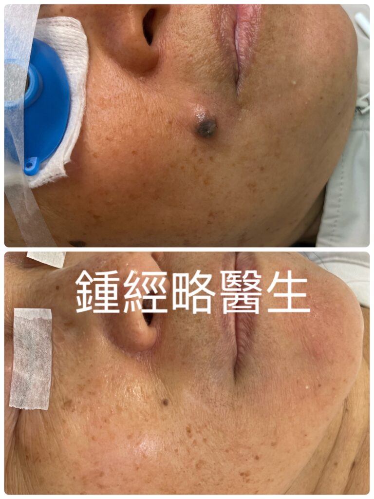
Location: Situated near the right lower nasolabial fold / perioral region, superior and lateral to the right oral commissure (corner of the mouth).
Lesion Type & Elevation: A raised, nodular/papular cutaneous lesion with a clear exophytic component protruding above the surrounding skin plane.
Coloration: Darkly pigmented, predominantly dark brown to blackish, displaying mild color heterogeneity across its surface.
Borders & Shape: Roughly circular to oval with moderately well-defined, somewhat rounded borders.
Surface Characteristics: The surface appears smooth to slightly lobulated with a faint shiny/reflective highlight on its superior aspect.
Surrounding Skin: The adjacent facial skin shows fine pore structure, faint freckling/lentigines, and no marked surrounding erythema (redness) immediately around the base.
Impression: This is a benign compound skin nevus with deep-seated melanocyte nests (mole roots).
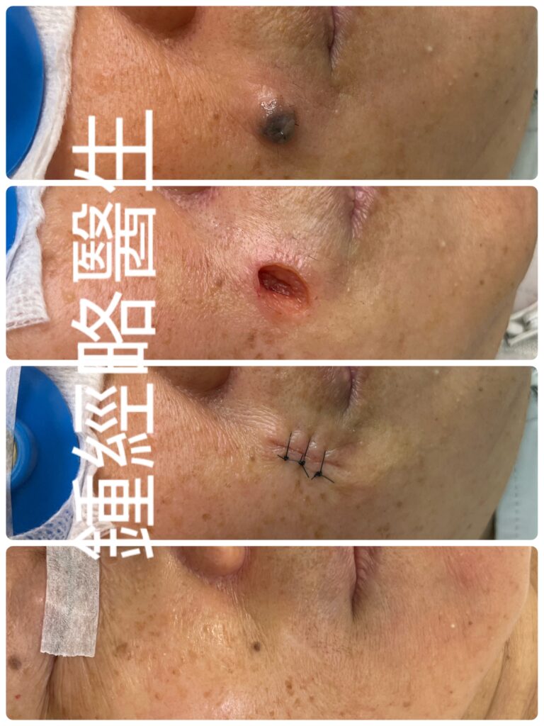
Treatment Performed: First, I precisely ablated the skin mole using a CO₂ laser to vaporize all melanocyte nests and prevent recurrence. Next, I reshaped the wound into an ellipse, which effectively prevents dog-ear deformity after closure. Finally, I sutured the wound layer by layer to ensure proper healing.
醫療報告
- 位置: 病灶部位鄰近右側下鼻唇溝及口周區域。具體而言,它部位於右侧口角(嘴角)上方偏外側。
- 病灶類型與隆起程度: 這是凸起的結節狀或丘疹狀皮膚病灶。此外,它含有明顯向外生長的部分,高於周圍皮膚表面。
- 顏色: 病灶色素沉著較深,主要呈現深棕色至黑褐色。而且,它的表面呈現輕微的顏色不均勻現象。
- 邊緣與形狀: 外觀大致呈圓形至橢圓形。再者,它具有中度清晰且偏圓潤的邊緣。
- 表面特徵: 整體而言,表面顯得平滑或呈輕微分葉狀。因此,其上方呈現微弱的光澤與反射光斑。
- 周圍皮膚: 與此同時,鄰近的面部皮膚顯示出細緻的毛孔與淡色雀斑。然而,根部周圍並沒有明顯發紅的現象。
- 診斷印象: 總結來說,這是良性的複合型皮膚痣,含有位置較深的黑色素細胞巢(痣根)。
- 實施治療: 首先,我使用二氧化碳雷射精準氣化這顆痣。如此一來,這便能清除所有黑色素細胞巢並防止復發。接著,我將傷口修整為橢圓形。因此,這能有效預防縫合後出現犬耳狀畸形。最後,我進行分層縫合,從而確保傷口順利癒合。
Discussion: Surgical Precision on the Nasolabial Fold: Navigating Complications, Scarring, and Recovery in Mole Excision
The nasolabial fold (NLF)—stretching from the nasal ala to the corner of the mouth—presents a high-stakes challenge for facial surgeons. While moles in this zone are common, removing them requires extreme caution. The fold hides a dense network of blood vessels, nerves, and lymphatic channels right beneath a dynamic, mobile surface. Consequently, executing a clean excision demands sound anatomical knowledge and precise surgical technique.
1. Surgical Challenges and Structural Risks
The soft tissue of the nasolabial fold consists of delicate, interconnected layers. A clear understanding of these sub-surface structures helps prevent inadvertent damage during excision.
       [ Superficial / Dermal Layer ]
                      |
        ( Facial Artery & Vein )
                      |
   [ SMAS / Facial Muscles (zygomaticus, levator) ]
                      |
  ( Infraorbital N. / Facial N. Marginal Br. )
Critical Structures at Risk
- Vascular Structures (Facial Artery and Vein): The facial artery runs deep beneath the facial muscles before emerging superficially near the nose as the angular artery. Cutting or nicking this vessel causes rapid bleeding, hematoma formation, and localized tissue death. Furthermore, post-operative hematomas increase wound tension and elevate infection risks.
- Nerve Branches (Sensory and Motor):
- Infraorbital Nerve: This nerve exits just below the lower eye socket. Transecting or compressing it causes persistent numbness, tingling, or burning sensations across the upper lip and cheek.
- Facial Nerve Motor Branches: These branches control facial expressions. Severing them causes lip drooping, asymmetry, and visible weakness during smiling or speaking.
- Lymphatic Channels: Transverse incisions across the natural drainage pathways disrupt fluid flow. As a result, patients experience prolonged swelling, tissue bogginess, and firm scarring along the upper lip and cheek.
2. Intraoperative Strategies to Avoid Complications
Surgeons can avoid structural injuries by adopting refined techniques that safeguard deeper planes.
Essential Surgical Rules
- Align with Natural Creases: Map the excision ellipse directly within the relaxed skin tension lines of the fold. Maintain a 3:1 or 4:1 length-to-width ratio to avoid unsightly “dog-ear” skin puckering at the tips.
- Control Depth and Undermining: Perform dissection strictly within the upper subcutaneous fat layer. Stay superficial to the muscle fascia to shield underlying nerve trunks. When releasing skin edges, keep undermining gentle, shallow, and parallel to the surface.
- Control Wound Depth, Width, and Bleeding Cleanly Use CO₂ laser ablation in Ultra-pulse mode layer by layer instead of surgical excision. Consequently, this approach seals small bleeding points without spreading heat to surrounding structures. Furthermore, the laser prevents char formation, so you can clearly visualize the underlying tissue. As a result, you maintain precise control over wound depth and avoid removing extra tissue. Additionally, local anesthetics with epinephrine (1:100,000) minimize capillary bleeding to keep the anatomical field clear.
3. Expediting Post-Operative Recovery
Fast, uncomplicated recovery depends on active wound care, inflammation control, and structural protection.
| Recovery Phase | Primary Goal | Clinical Actions |
| Immediate (0–48 hrs) | Prevent Hematoma & Edema | Apply compresses for a few hours. Elevate the head. Avoid bending or heavy lifting. |
| Early Wound (Days 2–7) | Support Re-Epithelialization | Clean gently with mild antiseptics. Can use alcohol after a few days. Keep the wound dry to prevent infection. |
| Suture Removal | Reduce Track Marks | Remove surface skin sutures at 10-14 days |
| Maturation (Weeks 2–12) | Foster Tissue Remodeling | Resume normal facial expressions gradually. Apply broad-spectrum sunblock to stop hyperpigmentation. |
4. Minimizing Scar Formation
Hiding a scar inside a natural facial crease requires zero surface tension and diligent post-operative management.
Surgical Closure Techniques
Place buried, absorbable sutures (5-0) deep within the dermal layer to absorb all underlying pull. Once the deep layer holds the weight, approximate the outer skin edges with fine, non-absorbable sutures (5-0) with minimal tension. Evert the epidermal edges slightly during closure when indicated.
Non-Invasive Scar Management
- Silicone Gel: Begin applying silicone few days after suture removal. Daily use for 4 to 6 months calms overactive collagen production and keeps the scar flat.
- Strict Sun Protection: Shield fresh tissue from sunlight to prevent post-inflammatory hyperpigmentation. Apply chemical and physical sunscreen daily for at least 6 months.
- Control Facial Movement: Limit excessive smiling, laughing, or chewing during the first two weeks. In high-mobility areas, small micro-botulinum toxin injections near the fold can temporarily immobilize the muscle and ease tension on the healing scar.
- Vascular Laser Therapy: If redness or prominent capillaries persist beyond 6 months, target the micro-vessels with a vascular laser to fade the mark.
醫學討論:鼻唇溝手術的精準手藝:如何應對切除痣時的併發症、疤痕與恢復
鼻唇溝(NLF)從鼻翼一路延伸至嘴角。這區塊對面部外科醫生而言,無疑是一項極高風險的挑戰。雖然這個區域的痣相當常見,但切除過程必須極度謹慎。畢竟,溝紋下方覆蓋著動態且富彈性的皮膚,底層更隱藏著密集的血管、神經與淋巴網絡。因此,執行乾淨俐落的切除手術,不僅需要紮實的解剖學知識,更仰賴精準的手術技巧。
1. 手術挑戰與結構風險
鼻唇溝的軟組織由非常精細且相互連結的層次組成。因此,清晰掌握這些皮下結構,能有效預防切除過程中的意外損傷。
       [ 表皮 / 真皮層 ]
               |
        ( 面動脈與面靜脈 )
               |
   [ SMAS 筋膜 / 面部肌肉(顴肌、提上唇肌)]
               |
   ( 眶下神經 / 面神經邊緣下頜支 )
面臨風險的關鍵結構
- 血管結構(面動脈與面靜脈): 面動脈走形於面部肌肉深層,接著在靠近鼻子處穿出至淺層,形成眶角動脈。倘若割傷或劃破這條血管,會迅速引發大量出血、血腫甚至局部組織壞死。此外,術後血腫更會增加傷口張力,進而提高感染風險。
- 神經支(感覺與運動):
- 眶下神經: 這條神經正好從眼眶下緣下方穿出。一旦切斷或壓迫它,會導致上唇與臉頰出現持續性的麻木、刺痛或灼熱感。
- 面神經運動支: 這些分支負責控制面部表情。若是切斷它們,會造成嘴唇下垂、兩側不對稱,以及在微笑或說話時出現明顯的面部無力。
- 淋巴通道: 如果橫向切斷自然的淋巴回流路徑,便會破壞體液循環。結果,患者的上唇與臉頰會面臨長期腫脹、組織水腫以及硬化疤痕。
2. 預防併發症的術中策略
外科醫生可以透過精細的操作技巧來保護深層結構,從而避免損傷重要組織。
手術核心原則
- 順應自然皮紋: 首先,請將預計切除的紡錘形(橢圓形)切口,精準定位於鼻唇溝的鬆弛皮膚張力線內。同時,請保持長寬比為 3:1 或 4:1。這樣一來,便能防止切口兩端出現難看的「犬耳狀」皮膚隆起。
- 控制深度與皮下剝離: 再者,請嚴格將剝離範圍限制在上層皮下脂肪段。保持在肌肉筋膜淺層操作,如此便能保護下方的神經主幹。當你釋放皮膚邊緣時,請確保剝離動作輕柔、淺層,並與皮膚表面保持平行。
- 乾淨控制傷口深度、寬度與止血: 請使用超脈衝模式的二氧化碳雷射(CO₂ laser)進行分層氣化,以替代傳統的手術刀切除。因此,這個方法可以在不將熱能擴散至周圍組織的情況下,迅速封閉微血管出血點。此外,雷射能防止焦炭形成,讓你能清晰觀察下方組織。結果,你便能精準掌控傷口深度,避免切除過多無辜組織。另外,配合使用含有腎上腺素(1:100,000)的局部麻醉劑,更能減少毛細血管出血,保持解剖視野清爽。
3. 加速術後恢復
想要達到快速且無併發症的恢復,關鍵在於積極的傷口照護、發炎控制以及結構保護。
| 恢復階段 | 主要目標 | 臨床處置 |
| 即刻期(0–48 小時) | 預防血腫與水腫 | 冰敷數小時。適度抬高頭部。同時,請避免彎腰或提重物。 |
| 傷口早期(第 2–7 天) | 促進表皮再生 | 使用溫和的消毒劑輕柔清潔。幾天後可以使用酒精消毒。請保持傷口乾燥以防感染。 |
| 拆線期 | 減少縫線痕跡 | 在第 10 至 14 天時拆除表皮縫線。 |
| 成熟期(第 2–12 週) | 促進組織重塑 | 逐步恢復正常的面部表情。塗抹廣譜防曬霜以阻絕色素沉著。 |
4. 降低疤痕形成
若想將疤痕完美隱藏於自然的面部褶皺中,必須做到表面零張力,並配合術後嚴格的管理。
手術縫合技巧
首先,請在真皮深層放置可吸收的可吸收縫線(5-0),藉此吸收底層所有的拉力。一旦深層結構承載了主要拉力,接著再使用細緻的不可吸收縫線(5-0),以極低的張力對齊外層皮膚邊緣。縫合時,若臨床情況允許,可將表皮邊緣微微外翻。
非侵入性疤痕管理
- 矽膠軟膏: 拆線幾天後,便可開始塗抹矽膠軟膏。每天持續使用 4 到 6 個月,這樣能平抑過度活躍的膠原蛋白增生,並讓疤痕保持平整。
- 嚴格防曬: 務必保護新生的肌膚免受陽光照射,進而預防發炎後色素沉著(PIH)。請每天塗抹含有物理與化學成分的防曬霜,並至少堅持 6 個月。
- 控制面部動作: 在前兩週內,請盡量減少過度的微笑、大笑或大聲咀嚼。針對高動態區域,可以在鼻唇溝附近施打微量肉毒桿菌素(Micro-botox),這能暫時放鬆肌肉,進而減輕復原中疤痕的張力。
- 血管雷射治療: 如果術後 6 個月以上依然殘留紅斑或明顯的毛細血管,可使用血管雷射精準擊退微血管,從而使痕跡淡化。