【注意:本文章及真實病人照片均源自鍾經略醫生的臨床皮膚科碩士及美容醫學碩士課程案例,屬於原創醫學文獻。因此,本院嚴格保留所有版權。同時,我們禁止任何形式的轉載與複製。】
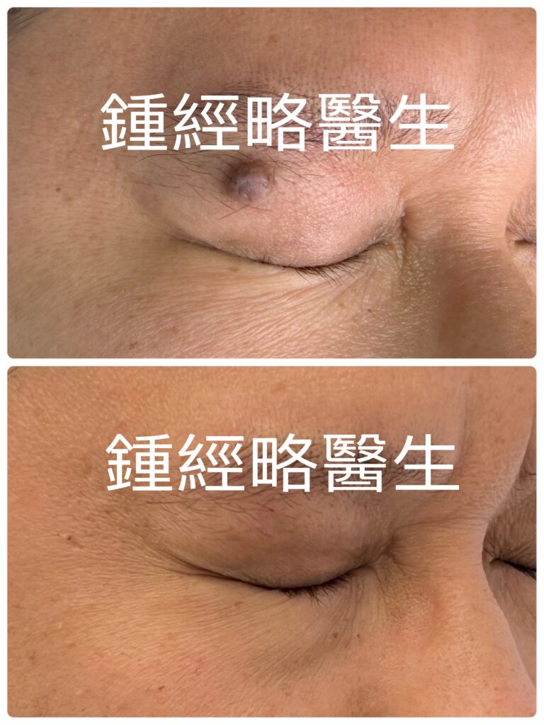
Above: Before and after picture of CO2 laser removal of a skin mole on left upper eyelid. The scar was almost unnoticeable several months post-op.
上圖:CO2 激光脫癦的手術前後對照相片。術後數個月後,患處幾乎看不見疤痕。
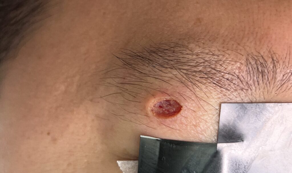
Above: After CO2 ablation of the skin mole, a depressed wound was senn
上圖:CO2 激光脫癦後可見一下陷的傷口。
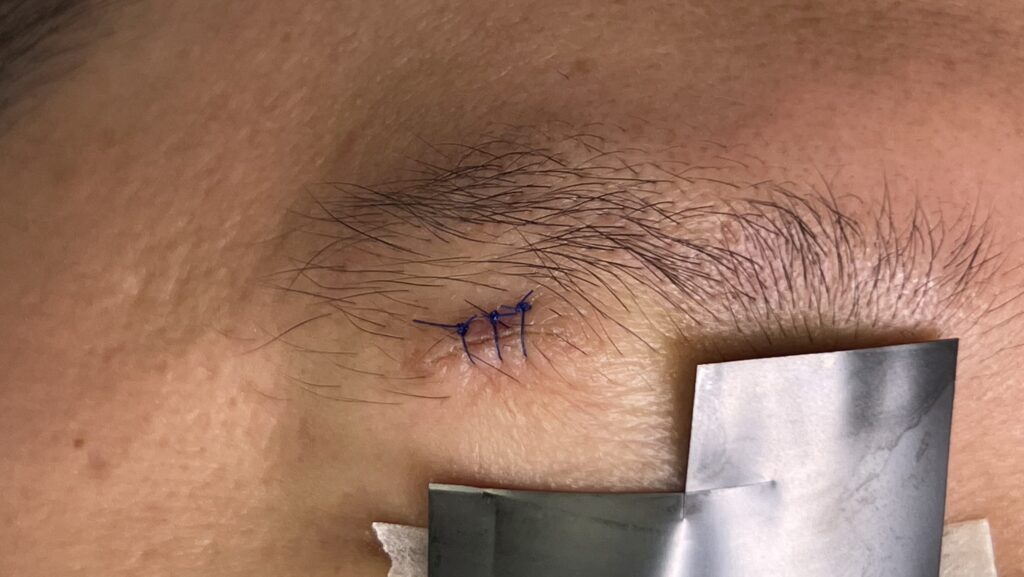
Above: The appearance of the wound after CO2 laser was sutured with 5-O nylon
上圖:激光脫癦後的傷口以5號尼龍線縫合
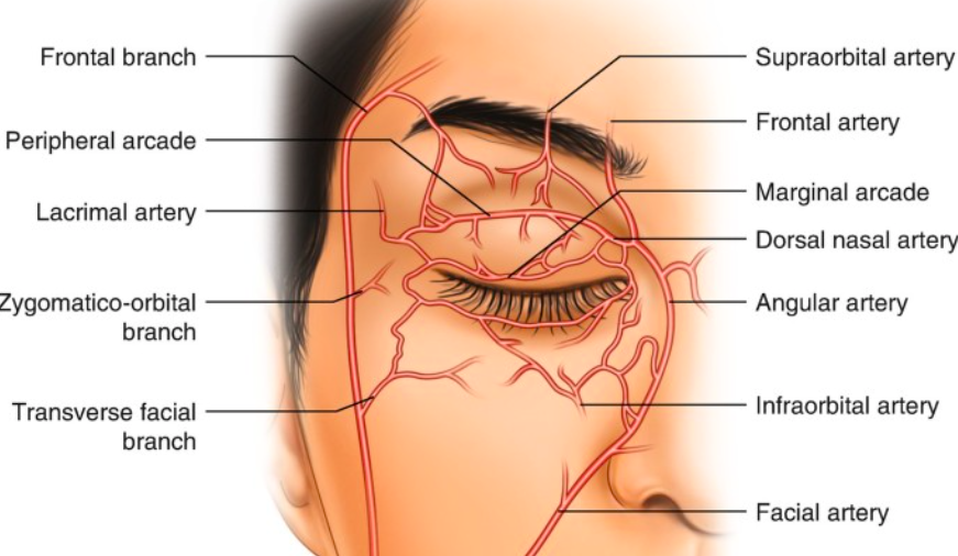
Above: Arteries of the upper eyelid
上圖:上眼皮的動脈分布圖
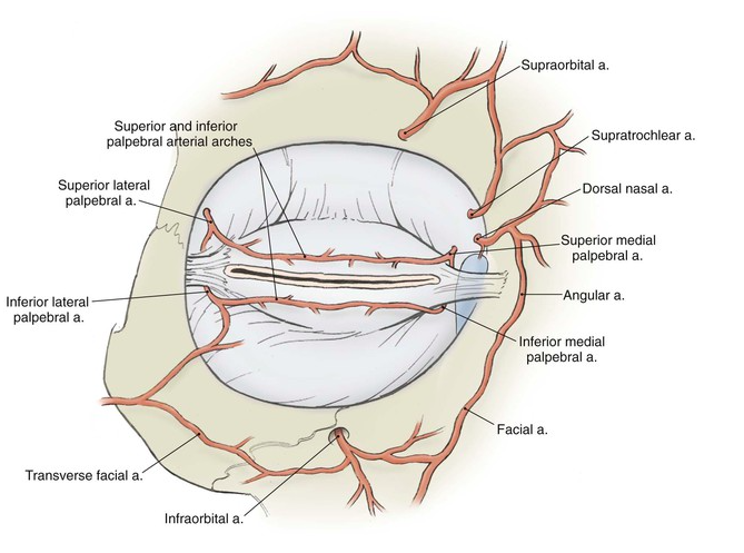
Above: Arteries of the upper eyelid
上圖:上眼皮的動脈分布圖
摘要
清除上眼瞼良性真皮黑色素痣/癦(Dermal melanocytic nevi),必須在「完整清除細胞」與「最小化結構變形」之間取得平衡。雖然傳統手術切除仍是標準選擇之一,但它通常需要較寬的安全邊緣、廣泛的組織剝離(Undermining),以及較長的線性切口以防止「犬耳」(Dog-ear)畸形。相反地,CO₂ 激光超脈衝模式(Ultrapulse mode) 的高精準度氣化能提供即時的光熱凝血、優異的深度控制,以及無碳化焦痂(Char-free)的組織氣化。因此,臨床醫生能在保護周圍健康組織的同時,直視並清除深層黑色素細胞巢(Melanocytic nests)。
本案例討論一例位於 50 歲男性、直徑為 0.8 cm 且高出皮膚表面 0.5 cm 的凸起真皮痣清除過程。透過結合超脈衝 CO₂ 激光氣化與標靶式一期縫合(Primary closure),我們成功實現了病灶完全清除、最小化切口長度,並達到理想的美學預後。
案例呈現
患者背景與病史
一名 55 歲男性因右上眼瞼長期存在凸起皮膚病灶前來求診。患者無顯著既往病史,亦無皮膚惡性腫瘤之家族病史。
- 病程時間: 病灶已存在超過 40 年,期間大小、形狀及顏色均無顯著改變。
- 臨床症狀: 患者完全無疼痛、瘙癢、壓痛、自發性出血或潰瘍。
- 主訴需求: 出於純粹的美觀考量要求去除病灶。
理學檢查與術前評估
於臨床檢查期間,我們觀察到:
- 病灶形態: 一個邊界清晰、淺褐色、無毛髮、高出皮膚表面的丘疹,測量大小為直徑 0.8 cm,高度 0.5 cm。
- 皮膚鏡檢查: 病灶展現均勻的色素分佈與良性真皮痣(癦)的結構,未見任何惡性變化的徵象。
- 解剖考量: 該痣部位於右上眼瞼皮膚,安全避開了下方主要的動脈幹線。
- 鬆弛度與張力測繪: 透過輕捏上眼瞼皮膚,我們觀察到顯著的年齡相關皮膚鬆弛度。此外,測繪放鬆皮膚張力線(RSTLs)後,確認採用水平方向進行切口最為合適。
手術處置:CO₂ 激光氣化與分層一期縫合
在與患者討論治療方案後,我們選擇了 CO₂ 超脈衝激光氣化,隨後進行一期縫合。此方案既滿足了他對極佳美學保留的需求,同時確保完全清除黑色素細胞的深層真皮巢。
                  [ 0.8 cm 凸起上眼瞼墨痣 ]
                                  │
             ┌────────────────────┴────────────────────┐
             ▼                                         ▼
  [ CO₂ 超脈衝激光 (Ultrapulse) ]             [ 一期分層縫合重建 ]
  • 精準逐層氣化病灶                         • 深層真皮 5-0 Monocryl 縫線
  • 直觀清除「根部」                           (無須剝離即可減張)
  • 無血、無碳化底座                         • 5-0 尼龍線進行表層精細對齊
1. 局部麻醉與激光氣化
首先,我們使用含有 1:100,000 腎上腺素的 2% 利多卡因(Lidocaine)進行局部麻醉,有效最小化術中微血管出血。接著,我們將 CO₂ 激光設定為 超脈衝模式(Ultrapulse mode)。由於超脈衝能在微秒級脈衝內釋放高能量,組織氣化速度極快,能防止熱量擴散至鄰近結構。
同時,我們在整個手術過程中使用冷生理鹽水持續沖洗周圍健康皮膚。這種主動冷卻保護了周邊組織免受附加的熱應力傷害。在 3× 放大鏡與無影手術燈下,我們對墨痣進行逐層氣化。
由於超脈衝模式能在不產生碳化焦痂的情況下封閉微血管,我們能清楚辨識黑色素細胞的深層真皮巢(墨痣的「根部」)。我們持續進行逐層氣化,直至徹底消除所有色素巢,留下一道乾淨、無血、凹陷的缺損,周圍為完好的正常真皮,且幾乎無熱傷害徵象。
2. 手術重建與縫合決策
在治療淺層癦痣(例如交界痣 Junctional nevi)時,臨床醫生可安全地讓小型缺損以二期癒合(Secondary intention)方式自行修復。然而,由於此真皮痣需要去除較深層的真皮組織,二期癒合會延長恢復期並增加疤痕攣縮的風險。因此,我們選擇了一期分層縫合。
傳統的手術教學建議採用 3:1 或 4:1 的紡錘形(Ellipse)長寬比,並進行廣泛的皮下剝離(Undermining)以避免皮膚產生「犬耳」起皺。然而,兩個關鍵的解剖因素改變了我們的策略:
- 原有的皮膚鬆弛度: 由於患者本身具備鬆弛的上眼瞼皮膚,我們安全地將切口比例縮減至 2.5:1,保留了大量健康皮膚。此外,也因為其鬆弛的上眼瞼皮膚,我們完全不需要進行皮下剝離。
- 無碳化的傷口床: 與傳統電灼(Electrocautery)會留下厚厚一層壞死組織與碳化焦痂不同,CO₂ 超脈衝激光氣化留下了乾淨、具活性(Viable)的傷口基底。因此,可以安全地使用縫合材料來對齊健康的組織邊緣。
我們使用 5-0 Monocryl 可吸收縫線 進行埋線深層真皮縫合以釋放機械張力,隨後使用 5-0 單絲尼龍線 進行單純間斷縫合,以實現精確的表皮對齊。
臨床結果與核心優勢
我們於術後 2 週拆除了表層尼龍線。拆線時,切口線平整柔軟,未見熱壞死、針眼痕跡或眼瞼緣變形。
總結:CO₂ 激光氣化相較於傳統冷鋼刀切除之優勢
- 更小的切口面積(Smaller Incisional Footprint): 激光氣化僅針對發生病變的組織。因此,臨床醫生能設計更短的紡錘形切口,無需不必要地切除健康組織。
- 卓越的深度控制與直視效果(Superior Depth Control & Direct Visualization): 由於超脈衝技術能在不產生焦痂的情況下實現即時止血,臨床醫生可在放大鏡下直接檢查真皮床,實時確認細胞已完全清除。
- 重建的靈活性(Reconstructive Versatility): 由於氣化邊緣保持活性且無焦痂,臨床醫生保留了隨即縫合缺損的完整靈活性,從而確保乾淨的一期癒合。
- 保護眼周血管網絡(Protection of Periorbital Vascular Networks): 精準的激光輸出將對上眼瞼精細微血管網絡的干擾降至最低,減少了術後瘀斑與水腫。
- 減少組織剝離的需求(Reduced Need for Tissue Undermining): 將激光精準度與患者現有的上眼瞼皮膚鬆弛度結合,既能縮短疤痕長度,又能避免侵入性的組織剝離。
- 快速且高質量的恢復(Rapid, High-Quality Recovery): 由於超脈衝氣化將附加熱傷害降至最低,傷口癒合速度與美學質量達到、且往往超過標準冷鋼刀切除的效果。
Abstract
Removing benign dermal melanocytic nevi from the upper eyelid demands an approach that balances thorough cellular clearance with minimal structural distortion. While traditional surgical excision remains a standard choice, it often requires wider safety margins, extensive tissue undermining, and longer linear incisions to prevent “dog-ear” deformities. Conversely, high-precision CO2 laser ablation in Ultrapulse mode provides immediate photothermal hemostasis, excellent depth control, and char-free tissue vaporization. Consequently, clinicians can directly visualize deep melanocytic nests while preserving healthy surrounding tissue.
This case discussion explores the removal of an 0.8cm (in diameter) elevated (0.5cm above skin level) dermal nevus on a 55-year-old male. By combining Ultrapulse CO2 laser vaporization with targeted primary closure, we achieved complete lesion eradication, minimal incisional length, and an optimal aesthetic outcome.
Case Presentation
Patient Profile & History
A 55-year-old male presented with a long-standing, elevated skin lesion on his right upper eyelid. He reported no significant past medical history or family history of dermatological malignancy.
- Duration: The lesion had been present for over 40 years without significant changes in size, shape, or color.
- Symptomatology: The patient experienced no pain, pruritus, tenderness, spontaneous bleeding, or ulceration.
- Chief Complaint: He requested removal solely for cosmetic improvement.
Physical Examination & Preoperative Assessment
During the clinical examination, we noted:
- Lesion Morphology: A well-demarcated, light brown, non-hirsute, elevated papule measuring 0.8cm in diameter and 0.5cm in elevation.
- Dermatoscopic Findings: The lesion exhibited uniform pigment distribution and a benign dermal nevus architecture, showing no signs of malignant transformation.
- Anatomical Considerations: The nevus occupied the right upper eyelid skin, situated safely away from major underlying arterial trunks.
- Laxity & Tension Mapping: By pinching the upper eyelid skin, we observed significant age-related skin laxity. Furthermore, mapping the relaxed skin tension lines (RSTLs) confirmed that a horizontal orientation would be most suitable.
Surgical Management: CO₂ Laser Ablation & Layered Primary Closure
After discussing the treatment options with the patient, we selected CO2 Ultrapulse laser ablation followed by primary suture closure. This approach met his desire for superior cosmetic preservation while ensuring complete removal of the deep intradermal nests of melanocytes.
                  [ 0.8 cm Elevated Upper Lid Nevus ]
                                  │
             ┌────────────────────┴────────────────────┐
             ▼                                         ▼
  [ CO₂ Laser Ultrapulse ]                 [ Primary Layered Closure ]
  • Precise layer-by-layer                 • Deep dermal 5-0 Monocryl
    photoevaporation                         (Tension relief without  undermining)
  • Direct visualization of roots          • Surface 5-0 nylon interrupted
  • Bloodless, char-free base                for fine epidermal alignment
1. Infiltration & Laser Vaporization
First, we administered local anesthesia using 2% lidocaine with 1:100,000 adrenaline, effectively minimizing intraoperative micro-bleeding. Next, we operated the CO2 laser in Ultrapulse mode. Because Ultrapulse delivers high energy in microsecond bursts, it vaporizes tissue fast and prevents heat from spreading to adjacent structures.
Simultaneously, we irrigated the surrounding healthy skin with cold normal saline throughout the procedure. This active cooling protected the peripheral tissue from collateral thermal stress. Working under 3 times magnifying loupes and shadowless operating lights, we vaporized the nevus layer by layer.
Because Ultrapulse mode seals micro-vessels without generating carbonized char, we could clearly identify the deep dermal nests (“roots”) of the melanocytes. We continued layer-by-layer ablation until we completely eliminated all pigmented nests, leaving a clean, bloodless, depressed defect bounded by pristine normal dermis with almost no signs of thermal damage.
2. Surgical Reconstruction & Closure Decisions
When treating shallow moles e.g. junctional nevi, clinicians can safely leave the small defect to heal by secondary intention. However, because this intradermal nevus required deeper dermal tissue removal, secondary intention healing would have prolonged recovery and increased the risk of scar contraction. Therefore, we chose primary layered closure.
Standard surgical teaching suggests using a 3:1 or 4:1 ellipse length-to-width ratio and performing extensive undermining to avoid “dog-ear” skin puckering. However, 2 key anatomical factors altered our strategy:
- Pre-Existing Skin Laxity: Because the patient possessed naturally loose upper eyelid skin, we safely reduced the incision ratio to 2.5:1, preserving substantial healthy skin. Also before of his loose upper eyelid skin, we eliminated the need for subcutaneous undermining
- Char-Free Wound Bed: Unlike traditional electrocautery, which leaves a thick layer of necrotic tissue and carbonised char tissue, Ultrapulse CO2 laser ablation leaves a clean, viable wound base. Consequently, suture materials can be safely used to oppose healthy tissue edges.
We placed buried deep dermal absorbable sutures using 5-0 Monocryl to relieve mechanical tension, followed by 5-0 monofilament nylon simple interrupted sutures for precise surface alignment.
Clinical Results & Key Advantages
We removed the surface nylon sutures 2 weeks postoperatively. At the time of removal, the incisional line appeared flat and soft with no signs of thermal necrosis, suture marks, or eyelid margin distortion.
Summary: CO₂ Laser Ablation vs. Traditional Cold-Knife Excision
- Smaller Incisional Footprint: Laser vaporization targets only the pathologically altered tissue. Therefore, clinicians can design a shorter ellipse without unnecessarily removing healthy tissue.
- Superior Depth Control & Direct Visualization: Because Ultrapulse technology achieves immediate hemostasis without creating char, clinicians can directly inspect the dermal bed under magnification and confirm complete cellular clearance in real time.
- Reconstructive Versatility: Because the ablated edges remain viable and uncharred, clinicians retain full flexibility to suture the defect immediately, securing clean primary intention healing.
- Protection of Periorbital Vascular Networks: Precise laser delivery minimizes disruption to the fine microvascular networks of the upper eyelid, reducing postoperative ecchymosis and edema.
- Reduced Need for Tissue Undermining: Combining laser precision with the patient’s existing upper lid skin laxity allows for shorter scars and avoids invasive undermining.
- Rapid, High-Quality Recovery: Because Ultrapulse ablation minimizes collateral thermal injury, wound healing matches—and frequently exceeds—the speed and cosmetic quality of standard cold-knife excision.