
【注意:本文章及真實病人照片均源自鍾經略醫生的臨床皮膚科碩士及美容醫學碩士課程案例,屬於原創醫學文獻。因此,本院嚴格保留所有版權。同時,我們禁止任何形式的轉載與複製。】
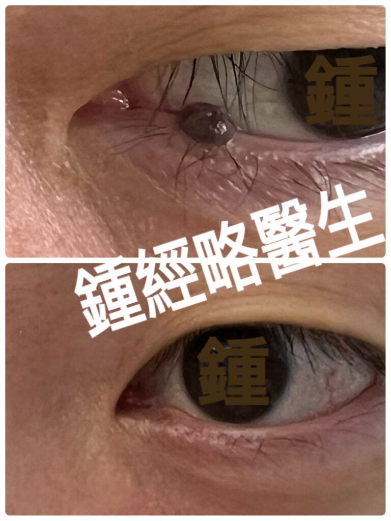
Before and after photograph of CO2 laser removal of a mole on left lower eyelid margin
左下眼瞼邊緣痣(眼線脫墨)二氧化碳激光脫墨術前術後照片
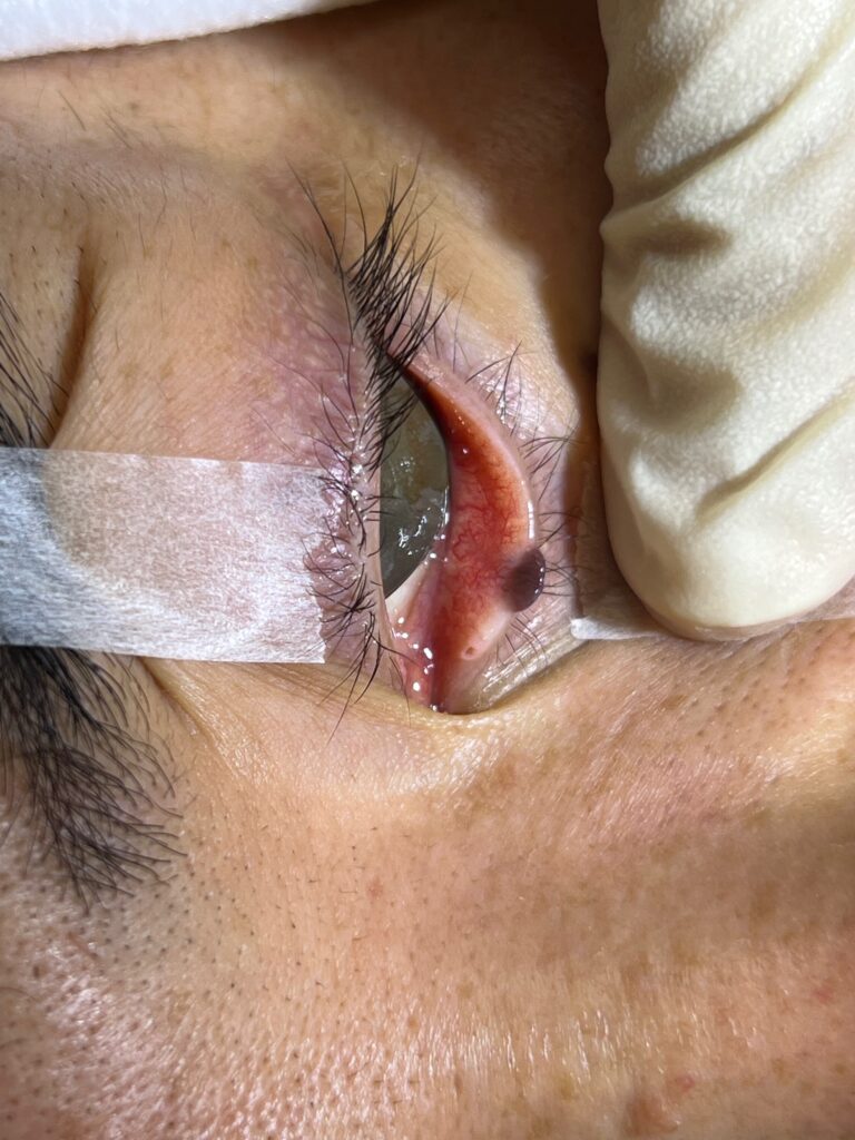
Preoperative photograph of CO₂ laser (eyelid margin / lash line) mole removal, demonstrating the deep extension of the nevus root, penetrating from the lower eyelid cutaneous surface through to the underlying palpebral conjunctival mucosa.
CO₂ 激光脫墨(眼瞼邊緣/眼線脫墨)之前的手術照片,顯示該墨痣的根部已經從下眼皮的皮膚滲透至該皮膚後的黏膜位置
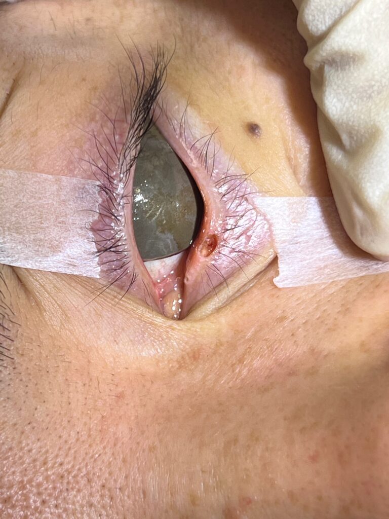
Intra‑operative photograph taken immediately after CO₂ laser ablation of lower eyelid margin skin mole, demonstrating a bloodless, char‑free skin wound extending to the reticular dermis
CO₂ 激光脫墨(眼瞼邊緣/眼線脫墨)之後的皮膚傷口(沒有流血及碳化,深度至真皮層)
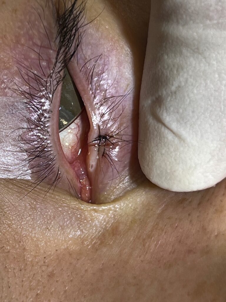
The eyelid margin wound was closed with 6-O nylon
眼瞼邊緣/眼線脫墨之後,傷口用6-O尼龍線縫合
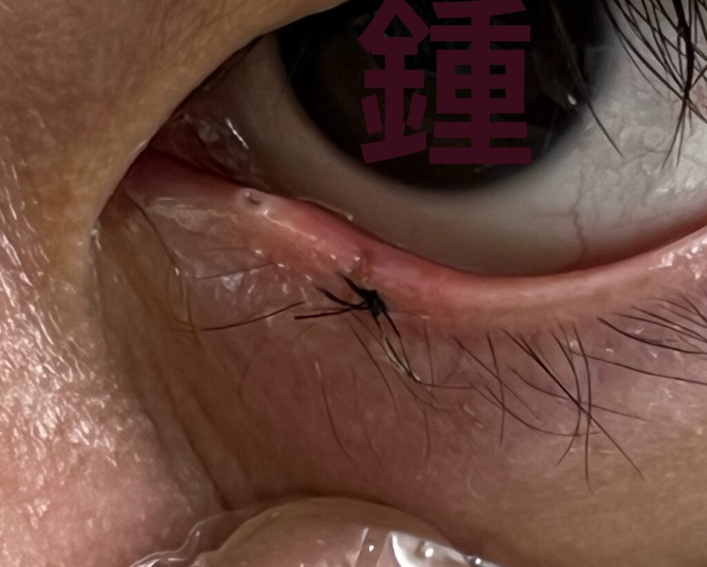
Postoperative photograph confirming optimal alignment of the eyelid margin. The smooth contour eliminates margin stepping, preventing secondary mechanical irritation to the cornea
術後照片顯示眼瞼邊緣已達到最佳對合。平滑的眼線輪廓(沒有瞼緣階梯狀不平)可避免對角膜造成次發性機械性刺激
Introduction
Large or protruding nevi along the ciliary margin can cause mechanical irritation, localized trichiasis, interference with the tear film distribution, and significant cosmetic concerns.
The management of melanocytic nevi located on the lower eyelid margin presents a delicate challenge in dermatologic and oculoplastic surgery. Because the eyelid margin is structurally complex—comprising ultra-thin skin, the orbicularis oculi muscle, the tarsal plate, Meibomian glands, and the ciliary margin contiguous with the palpebral conjunctiva—any intervention must balance complete lesion removal with the preservation of ocular function and cosmesis.
While traditional modalities like full-thickness wedge excision are historically utilized for eyelid lesions, they carry inherent risks of marginal notch deformity, lash loss, and prolonged healing times. Precision energy-based devices, such as carbon dioxide (CO2) lasers, offer a refined alternative.
Abstract
Melanocytic nevi on the eyelid margin present significant functional and aesthetic challenges in treatment due to their unique anatomical location. Traditional surgical excision often results in a full-thickness eyelid defect, requiring complex multi-layered suturing and easily leading to notch deformity and eyelash loss. This article reports a 50-year-old Chinese male patient who presented with a long-term, progressive mass on his left lower eyelid margin causing a foreign body sensation and visual obstruction. I used precise carbon dioxide (CO2) laser under local anesthesia. CO2 Laser ablation and microvaporization successfully eradicated deep melanocytes (the root of the nevus) without the need for pathological sections, while completely preserving the tarsal plate structure. During a 12-month follow-up, the patient showed no signs of recurrence, excellent functional recovery, and no noticeable scarring.
Clinical Presentation & History
The patient was a 50-year-old Chinese male with a history of good health and no major systemic diseases, ophthalmological history, or family history of skin cancer. He presented with a long-term, slowly growing skin lesion on his left lower eyelid margin, accompanied by intermittent foreign body sensation and partial visual obstruction.
Tracing the patient’s medical history, the lesion was first discovered when the patient was approximately 10 years old, initially as a small, flat macular nevus. Over the past forty years, the lesion has insidiously and slowly enlarged, gradually evolving into an exophytic, raised papule. The patient denies any recent signs of malignancy such as rapid enlargement, spontaneous bleeding, ulceration, severe itching, or pain.
Regarding functional effects, the patient reported that the pigmented nevus occasionally became congested and swollen, causing mechanical friction with the bulbar conjunctiva and cornea during blinking, resulting in corneal irritation and a foreign body sensation; in addition, the raised lesion caused partial visual field obstruction when gazing downwards into the peripheral field of vision.
Physical and Ophthalmological Examination
Examination of the ocular adnexa revealed a lesion located on the ciliary border of the left lower eyelid, presenting as a well-defined, dark brown, dome-shaped papule. The widest part of the lesion measured 3.0 mm, and it bulged outward from the eyelid margin by approximately 2.0 mm.
The dermoscopy and clinical details assessment are as follows:
- Symmetry and margins: The lesion presents as a bilaterally symmetrical oval structure with clear and distinct margins, and no irregular pseudopodia or extension to the palpebral conjunctiva is observed.
- Pigmentation and surface characteristics: It exhibits uniform dark brown pigmentation; the surface skin and mucous membranes are intact and smooth, without ulcers, hyperkeratosis or crusting.
- Surrounding tissue relationship: Adjacent eyelashes remain intact, with no localized eyelash loss (Madarosis). The lesion causes localized displacement of 1 to 2 adjacent Meibomian gland orifices, but the glands themselves do not show significant pathological changes.
Based on the above medical history and clinical manifestations, the diagnosis is:
- Benign Melanocytic Nevus
- Mechanical corneal and conjunctival irritation secondary to a mass on the eyelid margin.
- Mechanical visual obstruction secondary to eyelid margin lesions
Histological Decision
Given that the lesion was a skin pigmented nevus that had been present for decades and remained stable, with no recent morphological mutations, no family history of malignant tumors, and that the patient was of Chinese descent (the incidence of cutaneous malignant melanoma is extremely low), and the clinical assessment indicated a typical benign lesion. After discussion with the patient, it was determined that routine histopathological examination was not required in this case (Biopsy/Histology not required).
Surgical Approach and Postoperative Outcome
Given the lesion’s proximity to the sensitive and anatomically complex eyelash margin, I opted to administer precise carbon dioxide (CO2) under local anesthesia.
Surgical Procedure and Techniques
- Char-free Vaporization and Hemostasis: CO2 Laser treatment provides immediate coagulation of tiny blood vessels, resulting in minimal bleeding during the procedure. The clean, char-free surgical field allows surgeons to clearly observe the distribution of deep melanocytes and the nevus root within the skin tissue, enabling precise and complete removal while avoiding unnecessary excessive trauma.
- Deep debridement and protection of anatomical structures: During the procedure, microvaporization was used to completely remove melanocytes down to the reticular dermis. After the lesion was removed, a wound deep into the dermis was formed, but assessment confirmed that the underlying tarsal plate and orbicularis oculi muscle were intact.
- Simplified reconstruction and highly precise eyelid margin alignment: Compared to traditional surgery which requires addressing complex full-thickness defects, laser ablation leaves a relatively shallow wound, significantly simplifying the suturing process. To ensure the smoothness of the eyelid margin and prevent “stepping” that rubs against the cornea, I use 6-0 nylon sutures to precisely align and suture the wound from the deep dermis to the eyelid margin.
Postoperative follow-up
The patient’s postoperative recovery was smooth, with no complications such as infection or bleeding. The sutures were removed on the 12th day postoperatively. After 12 months of regular follow-up, the eyelid margin morphology recovered perfectly, no recurrence of the lesion was observed, and the wound left only a barely visible trace, achieving an excellent aesthetic and functional prognosis.
Discussion: Key advantages of laser treatment for removing pigmented nevi along the eyelid margin
The excision of eyelid margin lesions has always been a challenge in oculoplastic surgery. Compared to traditional surgical excision, high precision CO2 Laser ablation has demonstrated significant clinical and technological advantages:
1. Precisely locate the root of the mole to avoid excessive damage.
CO2 Laser allow surgeons to clearly visualize the root of the mole and the depth of the melanocytes, enabling complete removal without causing excessive trauma. In contrast, traditional surgical excision almost inevitably results in a full-thickness defect of the eyelid, forcing surgeons to perform complex multi-layered tissue reconstruction.
2. Significantly simplified repair procedure (Compared to Surgical Excision)
Traditional surgical resection requires sequential suturing and reconstruction of the tarsal plate, eyelid margin, and lower eyelid defects, which is technically demanding and time-consuming. CO2 Laser ablation usually leaves only a shallow dermal defect, making suturing simpler and easier; even in rare cases where it penetrates to form a full-thickness defect, the edge of the tarsal plate after laser ablation is clear and easy to identify and repair.
3. Superior hemostasis and clear visualization.
CO2 Laser possesses excellent microvascular instant coagulation capabilities, enabling a near-bloodless surgical field. This not only reduces intraoperative bleeding but also allows for clearer identification of tissue layers, ensuring a smoother suturing process.
4. Perfect preservation of eyelid anatomy and eyelashes (Preservation of Eyelid Architecture)
By calibrating appropriate laser parameters (ultra-pulse) and employing micro-layer vaporization, collateral thermal damage can be minimized. This selective vaporization technique preserves the natural contours of the eyelid margin and lash line, significantly reducing the risk of postoperative madarosis and notch deformities, while avoiding unnecessary trauma to adjacent ocular structures.
Conclusion
In summary, for benign melanocytic nevi located on the eyelid margin that are long-lasting, stable, and without any malignant potential, precise CO2 laser micro-layer ablation is recommended. CO2 Laser micro-layer ablation combined with deep suture offers multiple advantages, including complete removal of the mole root, excellent intraoperative hemostasis, simplified wound repair, and maximum preservation of eyelid structure. Compared to traditional full-thickness surgical excision, it is a minimally invasive preferred option that combines extremely high safety, functional protection, and excellent aesthetic outcomes.
前言
位於睫毛緣的巨大或凸起痣(墨)可能導致機械性刺激、局部倒睫、干擾淚膜分佈,並帶來顯著的美觀問題。
處理位於下眼瞼邊緣的色素痣(眼線脫墨),在皮膚科及眼整形外科領域是一項精細的挑戰。由於眼瞼邊緣結構複雜——包含極薄的皮膚、眼輪匝傷肌、瞼板、瞼板腺,以及與眼瞼結膜相連的睫毛緣——任何手術干預都必須在完整切除病灶與保留眼部功能及美觀之間取得平衡。
雖然傳統的「全層楔形切除術」在歷史上常用於眼瞼病灶,但其隨之而來的是眼瞼緣凹陷畸形、睫毛脫落及癒合時間延長等潛在風險。精準的能量設備,例如二氧化碳(CO₂)激光則提供了一個更精細的替代方案
摘要
眼瞼邊緣的黑色素痣(墨)因其特殊的解剖位置,在治療上存在顯著的功能與美觀挑戰。傳統外科切除常導致眼瞼全層缺損,需要複雜的多層縫合,並容易造成缺刻畸形及睫毛脫落。本文報告一例 50 歲中國男性患者,左下眼瞼邊緣長期進展性腫塊,導致異物感及視覺遮擋。我在局部麻醉下使用精準二氧化碳(CO₂)激光消融與微層汽化,成功徹底清除深層黑色素細胞(痣的根部),無需病理切片,並完全保留瞼板結構。術後隨訪 12 個月,患者無復發,功能恢復良好,且無明顯瘢痕。
臨床表現與病史
患者為 50 歲中國男性,身體健康,無重大全身疾病、眼科病史或皮膚癌家族史。主訴左下眼瞼邊緣長期、緩慢增大的皮膚痣(墨),伴間歇性異物感及部分視覺遮擋。
病史追溯至約 10 歲時,病灶初為小型扁平色素斑。四十年間逐漸隱匿性增大,演變為外生性隆起墨痣。患者否認近期惡性徵象,如快速增大、自發性出血、潰瘍、劇烈瘙癢或疼痛。
功能影響方面,患者報告痣偶爾充血腫脹,眨眼時與球結膜及角膜摩擦,引起角膜刺激與異物感;此外,隆起病灶在向下凝視時造成部分視野遮擋。
身體與眼科檢查
檢查顯示該墨痣位於左下眼瞼睫毛邊緣,呈界限清楚、深棕色、圓頂狀丘疹。最大直徑約 3.0 mm,自眼瞼邊緣向上隆起約 2.0 mm。
皮膚鏡與臨床特徵:
- 對稱性與邊界:病灶呈雙側對稱橢圓形,邊界清晰,無不規則假足或延伸至瞼結膜。
- 色素與表面特徵:均勻深棕色,表皮與黏膜完整光滑,無潰瘍、角化過度或痂皮。
- 周圍組織關係:鄰近睫毛完整,無局部脫落。病灶造成 1–2 個鄰近瞼板腺開口輕度移位,但腺體無顯著病變。
診斷:
- 良性黑色素痣(墨)
- 次發性角膜與結膜機械性刺激
- 次發性視覺遮擋
組織學決策
由於該墨痣存在數十年且穩定,無近期形態突變,無惡性腫瘤家族史,且患者為中國人(皮膚惡性黑色素瘤發病率極低),臨床評估為典型良性病灶,與患者商討後決定不作常規病理檢驗。
眼線脫墨之手術方式與術後結果
考慮病灶位於敏感且解剖複雜的睫毛邊緣,我選擇在局部麻醉下使用精準 CO₂ 激光進行眼線脫墨。
激光眼線脫墨手術技術:
- 無炭化汽化與止血: CO₂ 激光可即時凝固微血管,術中出血極少。清晰、無炭化的手術視野使醫生能清楚觀察深層黑色素細胞及痣根部,精確完整切除,避免不必要的創傷。
- 深層清除與解剖保護: 微層汽化徹底去除至網狀真皮層的黑色素細胞。術後形成真皮層深部傷口,但瞼板與眼輪匝肌保持完整。
- 簡化重建與精準瞼緣對位: 相較傳統手術需處理全層缺損,激光僅留下淺表傷口,顯著簡化縫合。為確保瞼緣平滑並避免角膜摩擦,我使用 6-0 尼龍線自真皮深層至瞼緣精準縫合。
激光眼線脫墨術後隨訪: 患者恢復順利,無感染或出血。術後第 12 天拆線。隨訪 12 個月,瞼緣形態恢復良好,墨痣無復發,傷口僅留極輕微痕跡,美容與功能效果皆佳。
討論:激光治療眼瞼邊緣色素痣的優勢
- 精確定位痣根,避免過度損傷 CO₂ 激光能清楚顯示痣根及黑色素細胞深度,完整切除而不造成過度創傷。傳統手術幾乎必然導致全層缺損,需複雜重建。
- 顯著簡化修復過程 傳統切除需依序縫合瞼板、瞼緣及下眼瞼缺損,技術要求高且耗時。激光消融通常僅留淺表缺損,縫合更簡單;即使少數形成全層缺損,瞼板邊緣亦清晰易於修復。
- 優越止血與清晰視野 CO₂ 激光具微血管即時凝固能力,術野幾乎無血,便於辨認組織層次,縫合更順利。
- 完美保留眼瞼解剖與睫毛 透過超脈衝與微層汽化,最大限度減少熱損傷。選擇性汽化技術保留瞼緣與睫毛線自然輪廓,顯著降低術後睫毛脫落與缺刻畸形風險,避免對鄰近眼部結構造成不必要創傷。
結論
總結而言,對於位於眼瞼邊緣的良性、長期穩定且無惡性潛能的黑色素痣,建議採用精準 CO₂ 激光微層消融。此技術結合深層縫合,具備多重優勢:徹底去除痣根、優異術中止血、簡化傷口修復、最大限度保留眼瞼結構。相較傳統全層外科切除,這是一種微創、極高安全性、兼具功能保護與美容效果的首選方案。