
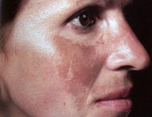
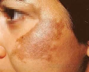
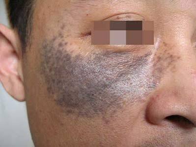
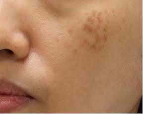
深層色素沉著(位於真皮層)通常呈灰棕色或藍灰色,比表皮色素更難治療,常見於黃褐斑的真皮型(荷爾蒙斑)、炎症後色素沉著(PIH)的深層型,以及一些先天性或後天性真皮黑色素沉著如太田痣、伊藤痣等。
常見的真皮層色素沉著類型
| 類型 | 深度 | 典型外觀 | 特點 |
|---|---|---|---|
| 真皮型黃褐斑(荷爾蒙斑) | 真皮層 | 灰棕或藍灰色斑塊,對稱分布於臉頰、額頭、上唇 | 與荷爾蒙、紫外線相關,慢性且易復發 |
| 深層炎症後色素沉著(PIH) | 真皮層 | 灰棕色斑痕,常見於痤瘡或濕疹後 | 在深膚色人群更常見,持續時間長 |
| 太田痣(Nevus of Ota)/ 伊藤痣(Nevus of Ito) | 真皮層 | 藍灰色斑塊,分布於三叉神經或肩臂區域 | 屬於真皮黑色素細胞增生,治療困難 |
| 苔癬樣色素沉著(Lichen Planus Pigmentosus) | 真皮層 | 灰棕色斑塊,常見於日曬或皺褶區域 | 慢性且難治療 |
| 接觸性皮膚炎後色素沉著(Riehl’s melanosis) | 真皮層 | 灰棕色斑塊,常見於化妝品或染料過敏後 | 屬於慢性接觸性皮膚炎的後果 |
臨床特徵
- 顏色:真皮層色素沉著通常呈灰棕或藍灰色,而表皮層則偏棕色。
- 治療反應:真皮層色素對光學治療(如 IPL、淺層雷射)反應差,需要更深層的 Q 開關 Nd:YAG 1064 nm ,皮秒激光,或長期藥物治療。
- 預後:治療困難,常需多次療程,且容易復發。
為何重要
區分表皮與真皮色素沉著是治療成功的關鍵。表皮型色素(如雀斑、日光性黑子)容易清除,而真皮型色素(如太田痣、真皮型黃褐斑/荷爾蒙斑)則更頑固,需要更長期且複合的治療策略。
✅ 總結:臉部的深層色素沉著包括 真皮型黃褐斑/荷爾蒙斑、深層 PIH、太田痣/伊藤痣、苔癬樣色素沉著、接觸性皮膚炎後色素沉著。這些病灶位於真皮層,顏色偏灰或藍灰,治療難度高,需謹慎選擇雷射波長與治療方式。
論文:皮秒激光/雷射在真皮層色素治療中的應用
引言
真皮層色素沉著因其深度、慢性特性及易復發而極難治療。傳統的 Q 開關雷射(532/1064 nm)長期以來是主要治療方式,但皮秒雷射——能在皮秒級釋放超短脈衝——已成為一種有前景的替代方案。它能更有效地擊碎色素顆粒,並減少熱損傷,特別適合頑固的真皮病灶。
作用機制
- 超短脈衝時間(皮秒級):產生強烈的光聲效應,將黑色素擊碎成比納秒/奈秒 Q 開關雷射更細小的顆粒。
- 降低熱擴散:減少對周圍組織的附帶損傷,降低疤痕及矛盾性加深(反黑)(paradoxical darkening)的風險。
- 多種波長可選:
- 1064 nm(紅外光):深入真皮,適合治療太田痣、伊藤痣及真皮型黃褐斑(荷爾蒙斑)。
- 532 nm(綠光):針對表淺色素,但在真皮病灶中 PIH 風險較高。
- 755 nm(亞歷山大光)與 730 nm:有時用於先天性真皮色素病灶。
臨床應用
- 真皮型黃褐斑:皮秒 1064 nm 分段模式可減淡灰棕斑塊,療程較少,但復發仍常見。
- 太田痣/伊藤痣:對皮秒 1064 nm 反應良好,通常需多次治療,但比 Q 開關清除速度更快。
- 深層炎症後色素沉著(PIH):皮秒雷射能更有效分解色素,減少痤瘡或濕疹後持久的色斑。
優點
- 效率更高:更細小的色素碎片更容易被巨噬細胞清除。
- 矛盾性加深風險較低:與 Q 開關 532 nm 相比,皮秒 1064 nm 對真皮色素更安全。
- 美容效果更佳:恢復期短、紅斑少、疤痕風險低。
- 患者舒適度高:研究顯示疼痛感較 Q 開關 Nd:YAG 低。
挑戰
- 復發:真皮型黃褐斑常因荷爾蒙及紫外線誘發而復發,與雷射種類無關。
- 成本高:皮秒設備價格遠高於 Q 開關雷射。
- 副作用:PIH 仍可能發生,尤其在深膚色患者中,雖然發生率略低於 Q 開關。
- 需多次療程:色素清除是漸進的,需事先告知患者合理期望。
結論
皮秒雷射(532/1064 nm)在治療真皮層色素方面是一項進步。其超短脈衝能更有效擊碎色素、減少熱損傷,並可能降低矛盾性加深的風險,相較於 Q 開關 Nd:YAG 更具優勢。然而,復發、成本及多次療程仍是挑戰。最佳療效需謹慎選擇波長、保守能量設定,並搭配嚴格的輔助措施,如防曬及外用淡斑藥物。
Deep pigmentation (located in the dermal layer) usually appears gray‑brown or bluish‑gray. It is more difficult to treat than epidermal pigmentation. It is commonly seen in dermal‑type melasma(hormone pigmentation), deep post‑inflammatory hyperpigmentation (PIH), and certain congenital or acquired dermal melanoses such as nevus of Ota and nevus of Ito.
Common Types of Dermal Pigmentation
| Type | Depth | Typical Appearance | Features |
|---|---|---|---|
| Dermal Melasma (Hormone pigmentation) | Dermis | Gray‑brown or bluish‑gray patches, symmetrically distributed on cheeks, forehead, upper lip | Related to hormones and UV exposure; chronic and prone to recurrence |
| Deep PIH (Post‑inflammatory Hyperpigmentation) | Dermis | Gray‑brown marks, often after acne or eczema | More common in darker skin types; long‑lasting |
| Nevus of Ota / Nevus of Ito | Dermis | Bluish‑gray patches, distributed along trigeminal nerve area or shoulder/arm region | Due to dermal melanocyte proliferation; difficult to treat |
| Lichen Planus Pigmentosus | Dermis | Gray‑brown patches, often in sun‑exposed or flexural areas | Chronic and resistant to treatment |
| Riehl’s Melanosis (Pigmentation after Contact Dermatitis) | Dermis | Gray‑brown patches, often after cosmetic or dye allergy | A consequence of chronic contact dermatitis |
Clinical Features
- Color: Dermal pigmentation usually appears gray‑brown or bluish‑gray, while epidermal pigmentation tends to be brown.
- Treatment Response: Dermal pigmentation responds poorly to optical therapies (e.g., IPL, superficial lasers). It requires deeper Q‑switched Nd:YAG 1064 nm treatment or long‑term medical therapy.
- Prognosis: Treatment is difficult, often requiring multiple sessions, and recurrence is common.
Why It Matters
Distinguishing epidermal from dermal pigmentation is critical for successful treatment. Epidermal pigmentation (such as freckles, solar lentigines) is easier to clear, while dermal pigmentation (such as nevus of Ota, dermal melasma/hormone pigmentation) is more stubborn and requires long‑term, combined treatment strategies.
✅ Summary: Deep facial pigmentation includes dermal melasma/hormone pigmentation, deep PIH, nevus of Ota/Ito, lichen planus pigmentosus, and Riehl’s melanosis. These lesions lie in the dermis, appear gray or bluish‑gray, are difficult to treat, and require careful selection of laser wavelength and treatment methods.
Essay: Picosecond Laser in the Treatment of Dermal Pigmentations
Introduction
Dermal pigmentations are notoriously difficult to treat due to their depth, chronicity, and tendency to relapse. Traditional Q‑switched lasers (532/1064 nm) have long been the mainstay, but picosecond lasers—delivering ultra‑short pulses in the picosecond range—have emerged as a promising alternative. Their ability to shatter pigment particles more efficiently with less thermal damage makes them particularly suitable for stubborn dermal lesions.
Mechanism of Action
- Ultra‑short pulse duration (picoseconds): Produces a strong photoacoustic effect, fragmenting melanin into finer particles than nanosecond Q‑switched lasers.
- Reduced thermal diffusion: Minimizes collateral damage to surrounding tissue, lowering risk of scarring and paradoxical darkening.
- Multiple wavelengths available:
- 1064 nm (infrared): Penetrates deeply into the dermis, ideal for nevus of Ota/Ito and dermal melasma/hormone pigmentation.
- 532 nm (green): Targets superficial pigment but carries higher risk of PIH in dermal lesions.
- 755 nm (alexandrite) and 730 nm: Sometimes used for congenital dermal pigment.
Clinical Applications
- Dermal Melasma/hormone pigmentation: Picosecond 1064 nm fractional mode can lighten gray‑brown patches with fewer sessions, though recurrence remains common.
- Nevus of Ota/Ito: Responds well to picosecond 1064 nm, often requiring multiple treatments but with faster clearance compared to Q‑switched.
- Deep PIH: Picosecond lasers can break down pigment more efficiently, reducing long‑lasting marks after acne or eczema.
Advantages
- Higher efficiency: Smaller pigment fragments are more easily cleared by macrophages.
- Lower risk of paradoxical darkening: Compared to Q‑switched 532 nm, picosecond 1064 nm is safer for dermal pigment.
- Better cosmetic outcomes: Reduced downtime, less erythema, and lower risk of scarring.
- Patient comfort: Studies show lower pain scores compared to Q‑switched Nd:YAG.
Challenges
- Recurrence: Dermal melasma/hormone pigmentation often relapses due to hormonal and UV triggers, regardless of laser type.
- Cost: Picosecond devices are significantly more expensive than Q‑switched lasers.
- Side effects: PIH still occurs, especially in darker skin types, though incidence may be slightly lower than with Q‑switched.
- Multiple sessions required: Clearance is gradual; patients must be counseled about realistic expectations.
Conclusion
Picosecond lasers (532/1064 nm) represent an advancement in the treatment of dermal pigmentations. Their ultra‑short pulses allow more efficient pigment fragmentation, reduced thermal damage, and potentially lower risk of paradoxical darkening compared to Q‑switched Nd:YAG. However, recurrence, cost, and the need for multiple sessions remain challenges. Optimal outcomes require careful wavelength selection, conservative fluence, and strict adjunctive measures such as sun protection and topical depigmenting agents.