醫學美容碩士及臨床皮膚科碩士雙碩士課程案例
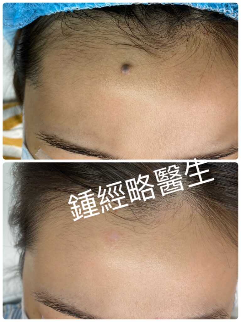
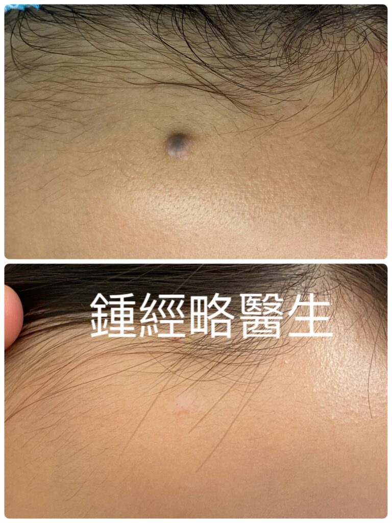
病患資料
年齡/性別: 25歲女性
過去病史: 無特殊病史
家族病史: 無特殊病史
現病史
患者自出生起,前額中央靠近髮際線處即有一處無症狀的皮膚病灶。該病灶在童年時期緩慢增大,但在她 15 歲時趨於穩定。先前的照片顯示為一個均勻、黑色且隆起的丘疹,帶有正常的皮膚紋理,內部無毛髮。她從未出現過疼痛、瘙癢、壓痛、發紅、腫脹、分泌物滲出或潰瘍。
兩年前,她出於美容考量尋求皮膚科治療。皮膚科醫生診斷為良性混合痣(benign compound nevus),並每隔兩個月進行一次二氧化碳(CO2)雷射表淺燒灼,共計八次。每次療程後,病灶隨後縮小和褪色出現暫時性結痂;然而,該病灶每次都會逐漸復發。因此,皮膚科醫生終止了雷射治療,並於六個月前將她轉診至整形外科。她隨後來到本診所,要求再次進行雷射治療,以避免手術切除並盡可能減少疤痕形成。
理學檢查
- 位置: 前額中央靠近前髮際線。
- 尺寸: 0.3 cm x 0.3 cm,高出周圍皮膚 0.2 cm。
- 形態: 邊界清晰、對稱的圓形丘疹。
- 表面/顏色: 缺乏正常皮膚紋理,且因反覆雷射熱損傷導致色素不均勻。
- 觸診: 無痛、質地軟、皮膚完整無潰爛且無分泌物。
臨床診斷
前額中央良性混合痣,因為前操作者的不良技術的關係,於多次不完全 CO2 雷射燒灼後復發。
尋找合適的醫生
一個同時擁有臨床皮膚科碩士及醫學美容碩士(雙碩士或以上)的醫生有能力擔任這個職責
手術過程與術中處置
手術項目: 放大鏡下高精度 CO2 雷射燒灼及分層一期縫合
手術步驟:
- 首先,具跨領域訓練的手術醫生(皮膚科訓練及美容醫學訓練)會先部分取樣並送檢(後證實為良性複合痣),隨後在 3.5 倍手術放大鏡下使用 UltraPulse CO2 雷射氣化深層色素細胞,同時保護周圍組織。
- 與此同時,團隊全程持續使用醫療級排煙機。
- 接著,具跨領域手術醫生經肉眼確認(以3.5倍手術放大鏡輔助)創面底部已完全清除深層色素細胞。
- 由此,團隊實現了完全止血,且無組織焦化或神經血管損傷。
- 最後,為確保最佳的美觀效果,手術醫生輕柔游離鄰近皮膚邊緣並進行分層縫合:深層組織使用 6-0 可吸收縫線對合,表皮則使用三針 5-0 尼龍線進行縫合。
術後病程與隨訪
- 拆線時間: 術後 2 週。
- 恢復狀況: 順利,無併發症之傷口癒合。
- 預後: 術後 6 個月複查時,手術部位已完全上皮化,痕跡幾乎不可見,且無痣復發的跡象。
Patient Profile
Age/Sex: 25-year-old female
Past Medical History: Unremarkable
Family History: Unremarkable
History of Present Illness
The patient had an asymptomatic skin lesion on her central forehead near the hairline from birth. The lesion grew slowly during childhood, but it stabilized when she turned 15. Prior photos showed a uniform, black, raised papule with normal skin lines and no hair inside. She never experienced pain, itching, tenderness, redness, swelling, fluid drainage, or open sores.
Two years ago, she sought skin care for cosmetic reasons. A dermatologist diagnosed a benign compound nevus and performed eight CO2 laser ablation sessions every two months. After each session, temporary crusts formed as the lesion shrank and faded. However, the spot gradually returned every time. Consequently, the dermatologist stopped laser treatments and referred her to plastic surgery six months ago. She then came to our clinic asking for another laser session to avoid surgery and minimize scarring.
Physical Examination
- Location: Mid-forehead next to the front hairline.
- Dimensions: 0.3 cm x 0.3 cm, raised 0.2 cm above the skin.
- Morphology: Clear borders, symmetrical, round papule.
- Surface/Color: Lacks normal skin lines with uneven coloring due to repeat laser heat damage.
- Palpation: Painless, soft, intact skin without fluid drainage.
Clinical Impression
Recurrent benign compound nevus of the central forehead following multiple incomplete CO2 laser ablations.
Finding the Right Doctor
A doctor with Dual Master Degrees in Clinical Dermatology and Aesthetic Medicine (or higher) is qualified to do this Cosmetic Operation.
Surgical Procedure & Intraoperative Management
Procedure: High-precision CO2 laser ablation and layered primary closure under magnification
Technique:
- First, the doctor with cross-disciplinary training did an incisional biopsy and sent specimen for histology examination to exclude malignancy (later confirmed to be a benign compound naevus). He then used an UltraPulse CO2 laser under 3.5x magnifying loupes to vaporize deep pigment cells while protecting surrounding tissue.
- Meanwhile, the team ran a medical-grade smoke evacuator continuously.
- Next, the doctor visually (with 3.5x surgical loupes) confirmed total removal of deep pigment cells (nests of melanocytes) at the wound base.
- Thus, the team achieved complete bleeding control without charring tissue or damaging nerves and blood vessels.
- Finally, to ensure the best cosmetic outcome, the doctor freed the nearby skin edges and stitched the wound in layers: absorbable 6-0 sutures for deep tissue, followed by three 5-0 nylon sutures for the top skin layer.
Postoperative Course & Follow-up
- Suture Removal: 2 weeks after surgery.
- Recovery: Smooth, complication-free wound healing.
- Outcome: At her 6-month checkup, the wound had fully healed with an almost invisible scar and no sign of the mole returning.