切除位於上唇(唇紅區域,vermilion)的皮膚痣,比起一般的皮膚切除術具有顯著的外科與解剖學挑戰。由於上唇是面部的視覺焦點,且具有獨特的組織特性,外科醫生必須克服複雜的美觀、功能與結構難關。
病史與主訴 (History & Presentation)
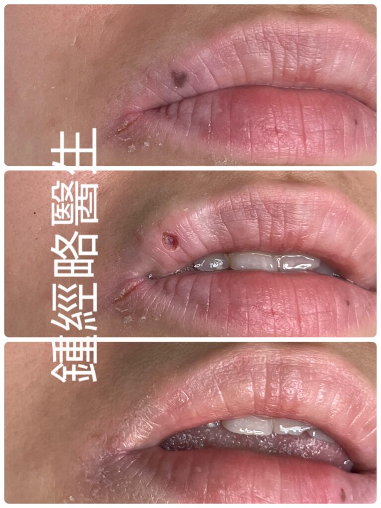
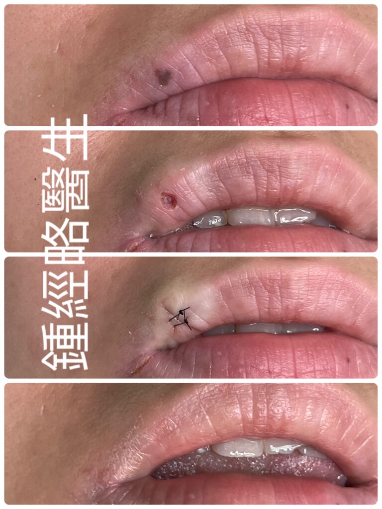
一名 37 歲女性,過去健康狀況良好且無顯著家族病史,因美觀考量求診,希望能切除唇部深褐色病灶。該位於右上唇紅緣的平坦小斑塊已有 20 年的極慢生長史,且在近 10 年內保持穩定。病灶邊緣不規則,顏色呈現均勻的深褐色。患者表示無疼痛、壓痛、發癢、潰瘍、出血、分泌物或結痂。
理學檢查 (Physical Examination)
檢查顯示病灶為 0.2 cm × 0.2 cm 的平坦深褐色斑塊,邊緣不規則,顏色均勻。觸診確認皮膚質地正常,無壓痛、發紅或腫脹。此外,病灶未見潰瘍、分泌物、出血或惡性徵象。局部淋巴結觸診未發現腫大。
手術過程與結果 (Surgical Procedure & Outcome)
醫生注射含腎上腺素的 2% Lidocaine 進行局部麻醉。在無影手術燈與 3.5 倍手術放大鏡下,使用 Ultrapulse CO2 雷射進行精準汽化切除。雷射完全清除所有黑色素細胞巢至正常組織,過中無焦碳形成或出血。
汽化後產生的傷口尺寸為 0.28 cm × 0.28 cm,深度為 0.35 cm。檢查傷口底部確認黑色素細胞已完全清除。醫生隨後使用 5-0 尼龍線縫合傷口,並於術後第 12 天拆線。術後 4 個月複查,患者的傷口疤痕已近乎不可見。
外科挑戰與 CO2 雷射的解決方案 (Surgical Challenges & How CO2 Laser can help to overcome them)
1. 高血管分布與出血風險
- 挑戰: 上唇由上唇動脈及其微血管網絡豐富供血。切開此處組織會帶來極高的術中出血與術後血腫風險。
- 雷射解決方案: CO2 雷射可在汽化過程中封閉微血管,提供即時止血效果。它還能清楚顯影較大的血管以避免誤傷。即使雷射意外觸及較大靜脈或動脈,暫時性的加壓止血即可迅速控制出血。最終的傷口縫合可達到即時且確切的止血。與手術刀切開相比,雷射汽化極少引起血腫。
2. 脆弱組織與唇紅邊界對齊
- 挑戰: 唇紅上皮缺乏角質層,非常薄且脆弱。表層縫線過緊容易撕裂黏膜。此外,即使唇紅邊界(Vermilion border)出現小於 0.5 mm 的錯位,也會導致明顯的「階梯狀變形」(Step-off deformity)。麻醉藥帶來的組織腫脹(Tumescence)亦可能扭曲此邊界。
- 雷射與技術解決方案: 在注射局部麻醉藥前,先用手術墨筆標記唇紅邊界以確保精準對齊。此外,將深層縫線錨定於口輪匝肌,可將機械張力從脆弱的黏膜表面轉移開。
3. 動態運動與疤痕收縮
- 挑戰: 說話、微笑與進食時口輪匝肌的持續運動會對傷口產生巨大的機械應力,增加疤痕變寬或傷口裂開的風險。不當的切除方向還可能導致唇緣外翻或扭曲唇珠(Cupid’s bow)。
- 雷射與技術解決方案: 順應天然的唇紋(Smoker’s lines)設計傷口,可使疤痕收縮的方向一致,防止嘴唇變形。主要縫合能減輕持續的機械拉扯,從而穩定傷口邊緣並促進癒合。
4. 潮濕、口腔污染與環境
- 挑戰: 唾液、口腔細菌、食物及液體會持續將黏膜傷口暴露於污染中,使傳統敷料的使用變得困難。
- 雷射與技術解決方案: 術後立即塗抹氯黴素軟膏(Chloramphenicol ointment)與凡士林,可建立保護性屏障,隔絕唾液與食物殘渣。這種濕性傷口癒合方法能軟化組織,減輕大笑、打噴嚏或打哈欠等面部表情運動時的張力。
Excision of a skin nevus (mole) located on the upper red lip—the vermilion—presents distinct surgical and anatomical challenges compared to typical cutaneous skin excisions. Because the upper lip is a high-impact facial focal point with unique tissue characteristics, surgeons must navigate complex aesthetic, functional, and structural hurdles.
History & Presentation
A 37-year-old woman with an unremarkable medical and family history presented for cosmetic removal of a dark brown lip lesion. The small, flat macule on her right upper vermilion lip grew very slowly over 20 years and remained stable for the last 10 years. The lesion showed an irregular border and homogeneous dark brown pigmentation. However, she reported no pain, tenderness, itchiness, ulceration, bleeding, discharge, or scabbing.
Physical Examination
Examination revealed a 0.2 cm × 0.2 cm flat, dark brown macule with an irregular border and homogeneous color. Palpation confirmed normal skin texture with no tenderness, redness, or swelling. Furthermore, the lesion showed no signs of ulceration, discharge, bleeding, or malignancy. Palpation of local lymph nodes revealed no lymphadenopathy.
Surgical Procedure & Outcome
The surgeon administered local anesthesia using 2% lidocaine with adrenaline. Operating under a shadowless surgical lamp and a 3.5× surgical loupe, they precisely ablated the lesion using an Ultrapulse CO2 laser. The laser completely removed all melanocyte nests down to normal tissue without causing charring or bleeding.
The ablation created a wound measuring 0.28 cm × 0.28 cm with a depth of 0.35 cm. Inspection of the wound base confirmed complete melanocyte clearance. The surgeon then closed the wound with 5-0 nylon sutures and removed the stitches on Post-Op Day 12. At the 4-month follow-up, the patient demonstrated an almost invisible scar.
Surgical Challenges & How CO2 Laser can help to overcome them
1. High Vascularity & Bleeding Risk
- Challenge: The superior labial artery and its microvascular network supply the upper lip. Cutting this tissue creates a high risk of intraoperative bleeding and postoperative hematomas.
- Laser Solution: The CO2 laser provides immediate hemostasis by sealing small blood vessels during ablation. It also allows clear visualization of larger vessels to avoid accidental injury. If the laser hits a larger vein or artery, temporary external pressure quickly controls the bleeding. Final wound suturing provides immediate definitive hemostasis. Unlike a scalpel incision, laser ablation rarely causes hematomas.
2. Delicate Tissue & Border Alignment
- Challenge: The vermilion epithelium lacks a keratinized layer, making it thin and fragile. Tight surface sutures can easily tear the mucosa. Additionally, even a sub-millimeter mismatch at the vermilion border causes a noticeable “step-off” deformity. Anesthetic tumescence can distort this boundary.
- Laser & Technique Solution: Marking the vermilion border with surgical ink before injecting local anesthetic ensures precise alignment. Furthermore, anchoring deep muscular sutures into the orbicularis oris transfers mechanical stress away from the delicate mucosal surface.
3. Dynamic Motion & Scar Contraction
- Challenge: Continuous orbicularis oris movement during speech, smiling, and eating places high mechanical stress on the wound, increasing the risk of scar widening or dehiscence. Improper excision orientation can also evert the lip margin or distort Cupid’s bow.
- Laser & Technique Solution: Designing the wound along natural smoker’s lines aligns the vector of scar contraction to prevent lip distortion. Primary suturing relieves continuous mechanical stretching, which stabilizes the wound edges and accelerates healing.
4. Moisture, Oral Contamination, & Environment
- Challenge: Saliva, oral bacteria, food, and fluids continuously expose the mucosal wound to contamination, complicating standard adhesive dressing use.
- Laser & Technique Solution: Applying Chloramphenicol ointment and Vaseline immediately after surgery creates a protective seal against saliva and food particles. This moist wound-healing approach softens the tissue and minimizes tension during facial movements such as laughing, yawning, or crying.