病歷與臨床表現:
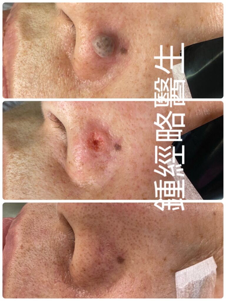
一名57歲男性因左側鼻翼長期存在皮膚病變前來就診。患者無特殊既往病史或家族病史。該病變最早出現於其6歲時,並在隨後的20年間緩慢增大;然而,數十年來其大小、形狀及顏色均保持完全穩定。患者提出切除要求純粹出於美觀考量。該均勻的淺褐色腫塊完全無症狀,無出血、潰瘍、瘙癢、疼痛或分泌物史。
體格檢查:
檢查顯示左側鼻翼有一大小約 0.6 × 0.6 cm 的穹窿狀淺褐色丘疹,高出皮膚表面 0.4 cm。色素分布均勻。此外,表面皮膚表現完全正常,保留了標準的皮膚紋理及皮膚附件/器官(包括毛囊口)。無出血、潰瘍、分泌物或結痂跡象,觸診時無壓痛。
臨床印象:
左側鼻翼良性皮內痣。
手術考量與挑戰:
於左側鼻翼進行手術具有顯著的解剖學挑戰。具體而言,該區域基礎血管分布較差、覆蓋皮膚較薄,且下方緊鄰鼻翼軟骨。此外,當地豐富的皮脂腺和汗腺持續分泌皮脂與汗液,在手術部位附近造成高風險的細菌污染環境。
手術過程:
- 麻醉與雷射氣化:首先,我們注射了 0.5 mL 不含腎上腺素的 2% 利多卡因進行局部麻醉。接著,我們使用超脈衝(Ultrapulse)模式的二氧化碳(CO₂)雷射精確氣化切除該痣。由於二氧化碳雷射能提供優異止血效果且能氣化組織而不致焦化,使我們能清晰觀察下方的真皮結構。這種清晰度有助於完全去除所有黑色素細胞巢以降低復發率,同時保留健康組織以防止疤痕形成。
- 切除與閉合:完全氣化後留下了一個較深的缺損。在此深度下,若僅靠二期癒合(secondary-intention healing)往往伴隨復原期延長,以及形成難看、凹陷且色素減退疤痕的高風險。因此,我們將缺損改為 3:1 的橢圓形切除,以防止「狗耳」(dog-ear)畸形並實現無張力縫合。在此處避免縫線張力至關重要;過度張力會損害/減少局部血液灌流,可能導致皮膚壞死,或在發生感染時大幅降低全身性抗生素的療效。
- 縫合:經精細的組織游離(undermining)後,我們使用可吸收縫線縫合皮下層。隨後,使用 5-0 尼龍縫線將皮膚邊緣精確對合。
術後照護與結果:
由於活躍的汗腺與皮脂分泌攜帶皮膚菌群,我們指示患者每日至少清潔傷口四次(若大量出汗或處於高溫環境中則需更頻繁)。術後恢復過程順利且無異常事件。我們於術後第 14 天拆除尼龍縫線。在術後 6 個月的隨訪中,手術部位癒合良好,疤痕幾乎不可見。
Patient History & Presentation
A 57-year-old male presented with a long-standing skin lesion on his left ala. He has no significant past medical history or family history. The lesion first appeared when he was 6 years old and grew slowly over 20 years. However, it has remained completely stable in size, shape, and color for decades. He requested removal strictly for cosmetic reasons. The homogeneous light brown lump was entirely asymptomatic, with no history of bleeding, ulceration, itching, pain, or discharge.
Physical Examination
Examination revealed a 0.6 × 0.6 cm dome-shaped, light-brown papule, raised 0.4 cm above the skin level. The pigment was uniform throughout. Additionally, the overlying skin appeared completely normal, retaining standard skin markings and cutaneous appendages (including follicular orifices). There was no evidence of blood, ulceration, discharge, or crusting, and the lesion was non-tender upon palpation.
Clinical Impression
Benign intradermal naevus of the left ala.
Surgical Considerations & Challenges
Operating on the left ala presents notable anatomical challenges. Specifically, this region features relatively poor baseline vascularity, thin overlying skin, and underlying alar cartilage. Moreover, abundant local sebaceous and sweat glands continuously secrete sebum and sweat, creating a high risk of bacterial contamination near the surgical site.
Surgical Procedure
- Anesthesia & Laser Ablation: First, we injected 0.5 mL of 2% lidocaine without adrenaline for local anesthesia. Next, we precisely ablated the naevus using a CO₂ laser in Ultrapulse mode. Because the CO₂ laser provides excellent hemostasis and vaporizes tissue without charring, we clearly visualized the underlying dermal structures. This clarity allowed complete removal of all melanocytic nests to minimize recurrence while simultaneously preserving healthy tissue to prevent scarring.
- Excision & Closure: Complete ablation left a deep defect. At this depth, primary secondary-intention healing often carries a high risk of prolonged recovery and unsightly, depressed, hypopigmented scars. Consequently, we converted the defect into a 3:1 elliptical excision to prevent dog-ear deformities and permit tension-free closure. Avoiding suture tension is critical here; excess tension impairs local blood perfusion, potentially leading to skin necrosis or severely reducing the efficacy of systemic antibiotics if an infection occurs.
- Suturing: After meticulous tissue undermining, we closed the subcutaneous layer using absorbable sutures. Subsequently, we accurately primary-opposed the skin edges with 5-0 nylon sutures.
Postoperative Care & Outcome
Because active sweat and sebaceous secretions harbor skin flora, we instructed the patient to cleanse the wound at least four times daily (and more frequently if sweating heavily or exposed to hot environments). The postoperative course was smooth and uneventful. We removed the nylon sutures on postoperative day 14. At the 6-month follow-up visit, the surgical site exhibited excellent healing with an almost unnoticeable scar.