額頭脫痣
病患資料與病史
a. 病患: 57 歲男性
b. 既往病史: 無特殊病史
c. 家族病史: 無特殊病史
d. 現病史: 病患因評估位於額頭中央、病程已久的先天性皮膚病灶而前來就診。該病灶首次出現於約 10 歲時,並緩慢生長至 20 歲。此後,其大小與外觀均保持穩定。病患表示無疼痛、壓痛、瘙癢、紅斑、水腫、潰瘍或分泌物。病灶上方未見多毛現象(hypertrichosis)。病患提出切除要求主要出於美觀考量。
身體檢查
a. 位置: 額頭中央
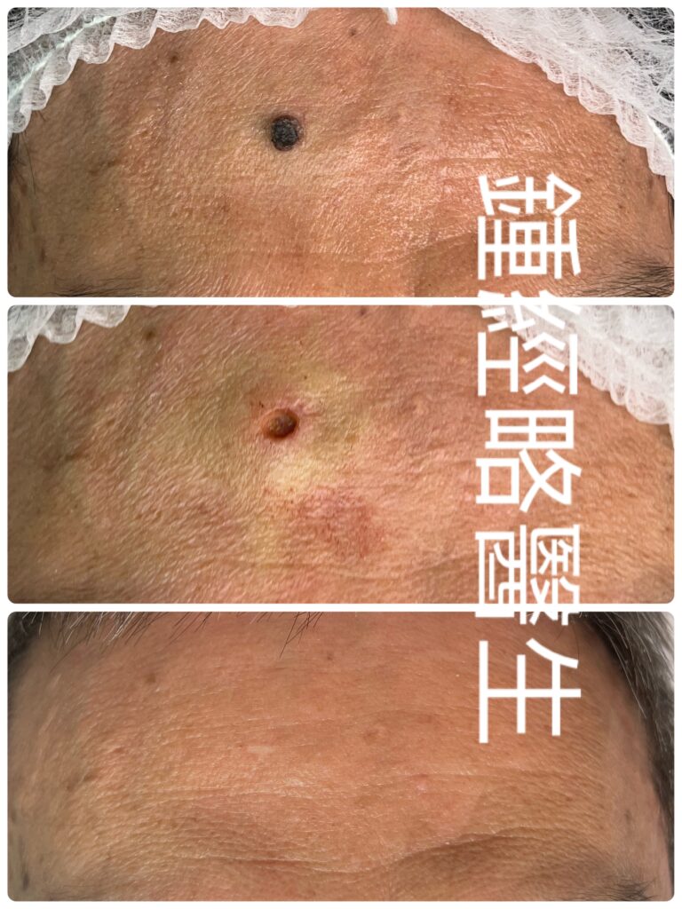
b. 型態: 對稱、大致呈圓形、均質深棕色丘疹
c. 邊緣: 界限清晰且規則
d. 尺寸: 0.3 公分 × 0.3 公分,較周圍皮膚表面隆起 0.2 公分
e. 表面特徵: 保留正常皮膚紋理;無紅斑、水腫、潰瘍或分泌物之跡象
f. 觸診: 無壓痛,具可動性
臨床印象
額頭中央良性混合痣(Compound nevus)。
手術過程細節
a. 麻醉: 使用含腎上腺素(1:100,000)之 2% Lidoacine 進行局部浸潤麻醉。
b. 汽化切除: 我們使用 UltraPulse CO2 激光對色素痣進行逐層汽化切除,深達真皮深層。此方法確保了所有深層黑色素細胞巢的完整清除。UltraPulse 模式發射的能量對鄰近組織的周邊熱損傷極小。此外,CO2 激光波長可實現同步止血且不產生焦炭(char)。因此,清晰的視覺反饋允許實時確認痣組織已完全清除至健康的真皮基質。未被汽化的基底保持了最佳的組織活力,有利於良好的傷口癒合。過程中未損傷任何重要血管或神經結構。
c. 重建與縫合: 鑒於所形成缺損的深度,我們進行了即時一期縫合,而非等待二期癒合:
- 皮下層: 使用 6-0 可吸收縫線進行內翻縫合,以減少深部無效腔(dead space)與傷口張力。
- 表皮層: 使用 5-0 尼龍線進行間斷縫合,以達到精確的皮緣對合。
術後過程與隨訪
a. 傷口護理: 病患每日進行無菌敷料更換,並嚴格避免浸水。
b. 拆線: 於術後第 12 天拆線,無併發症發生。
c. 6 個月隨訪: 手術部位展現出極佳的傷口成熟度,留下一條幾乎難以察覺的美觀疤痕,且無復發跡象。
臨床討論
皮膚外科中一個常見的誤解是:CO2 激光汽化切除與手術縫合是互斥的。傳統電灼或連續波激光會產生顯著的焦痂(eschar)與焦炭。這種僵硬的壞死組織會阻礙有效的組織游離與一期縫合。然而,超脈衝(ultra-pulsed)CO2 激光技術通過乾淨地汽化組織並將熱壞死降至最低,從而解決了這個問題。
某些良性痣(特別是混合痣和皮內痣)具有深層黑色素細胞巢。不完全的汽化切除存在高復發率的風險。相反,分段多次汽化切除往往因重複的機械與熱損傷而導致最終美觀效果變差。
通過將單次、完整的超脈衝 CO2 汽化切除與即時一期縫合相結合,我們獲得了關鍵的臨床優勢:
- 降低感染風險: 一期縫合消除了開放性肉芽傷口。
- 優異的美觀效果: 縫合防止了萎縮性或凹陷性疤痕的形成。
- 加速恢復: 一期癒合顯著縮短了總再表皮化(re-epithelialization)時間。
- 增強組織存活率: 極小的周邊熱損傷保留了皮緣活力,以實現無縫對合。
結論
使用高精度 CO2 UltraPulse 汽化切除聯合即時一期縫合,代表了一種在注重美觀的面部區域切除深層良性痣的高效、安全且具備卓越美觀效果的技術。
PATIENT DEMOGRAPHICS & HISTORY
- Patient: 57-year-old male
- Past Medical History: Unremarkable
- Family History: Unremarkable
- History of Present Illness: The patient presented for the evaluation of a long-standing congenital cutaneous lesion on the central forehead. The lesion first appeared around age 10 and grew slowly until age 20. It has remained stable in size and appearance since. The patient reports no pain, tenderness, pruritus, erythema, edema, ulceration, or discharge. No hypertrichosis is noted over the lesion. He requested excision primarily for cosmetic concerns.
PHYSICAL EXAMINATION
- Location: Central forehead
- Morphology: Symmetrical, roughly round, homogeneous deep brown papule
- Borders: Well-defined and regular
- Dimensions: 0.3 cm × 0.3 cm, elevated 0.2 cm above the surrounding skin surface
- Surface Features: Preserved normal skin markings; no evidence of erythema, edema, ulceration, or discharge
- Palpation: Non-tender, mobile
CLINICAL IMPRESSION
- Benign compound nevus, central forehead.
PROCEDURAL DETAILS
- Anesthesia: Local infiltration with 2% lidocaine with adrenaline (1:100,000).
- Ablation: We utilized an UltraPulse CO2 laser to ablate the nevus layer-by-layer down to the deep dermis. This approach ensured complete clearance of all deep-seated melanocytic nests.The UltraPulse mode delivers energy with minimal collateral thermal damage to adjacent tissues. Furthermore, the CO2 wavelength achieves simultaneous hemostasis without producing char. Consequently, clear visual feedback allowed real-time verification of complete nevus tissue clearance down to healthy dermal stroma. The non-vaporized base retained optimal tissue viability, favoring robust wound healing. No major vascular or neural structures were compromised.
- Reconstruction & Closure: Given the depth of the resulting defect, we performed immediate primary closure rather than allowing secondary intention healing:
- Subcuticular Layer: Inverted 6-0 absorbable sutures to reduce deep dead space and wound tension.
- Epidermal Layer: Interrupted 5-0 nylon sutures for precise skin edge apposition.
POSTOPERATIVE COURSE & FOLLOW-UP
- Wound Care: The patient performed daily sterile dressing changes and strictly avoided water immersion.
- Suture Removal: Sutures were removed on Postoperative Day 12 without complication.
- 6-Month Follow-Up: The surgical site demonstrated excellent wound maturation with an almost unnoticeable cosmetic scar and no signs of recurrence.
CLINICAL DISCUSSION
A common misconception in dermatologic surgery is that CO2 laser ablation and surgical suturing are mutually exclusive. Traditional electrocautery or continuous-wave lasers produce significant eschar and char. This rigid necrotized tissue prevents effective tissue mobilization and primary suturing. However, ultra-pulsed CO2 laser technology circumvents this issue by vaporizing tissue cleanly with minimal thermal necrosis.
Certain benign nevi, particularly compound and intradermal variants, feature deeply seated nests of melanocytes. Incomplete ablation risks high recurrence rates. Conversely, staged serial ablations often worsen final cosmesis due to repetitive mechanical and thermal trauma.
By combining single-stage, complete ultra-pulsed CO2 ablation with immediate primary closure, we achieved key clinical benefits:
- Reduced Infection Risk: Primary closure eliminates open granulating wounds.
- Superior Cosmesis: Suturing prevents the formation of atrophic or depressed scarring.
- Accelerated Recovery: Primary intention healing significantly shortens total re-epithelialization time.
- Enhanced Tissue Survival: Minimal collateral thermal injury preserves skin margin viability for seamless apposition.
CONCLUSION
High-precision CO2 UltraPulse ablation combined with immediate primary suturing represents an effective, safe, and cosmetically superior technique for removing deep benign nevi in cosmetically sensitive facial zones.