
【注意:本文章及真實病人照片均源自鍾經略醫生的臨床皮膚科碩士及美容醫學碩士課程案例,屬於原創醫學文獻。因此,本院嚴格保留所有版權。同時,我們禁止任何形式的轉載與複製。】
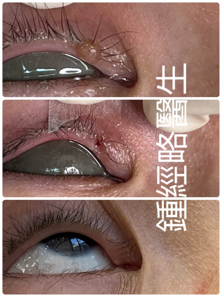
Case Presentation
Case Description
A 48-year-old female presented with a non-significant past health and family history. Her chief complaint was a skin-colored, benign mole on the medial side of her right upper eyelid margin. The lesion measured 0.3cm x 0.4cm, projected outward, and showed no ulceration, bleeding, or discharge. It grew slowly over 30 years without symptoms, and the patient requested laser removal for aesthetic reasons. Physical examination confirmed a benign mole that did not affect vision or scratch the cornea. The patient exhibited no other ocular signs or symptoms. Because the lesion was benign and the patient prioritized aesthetic outcomes, we chose CO2 laser removal without biopsy. This approach minimized wound size and eliminated the need for sutures.
Procedure & Results
Using 3.5x magnification loupes, we vaporized the mole and its underlying melanocytic nests with a CO2 laser to minimize recurrence. Immediately post-procedure, the wound showed a slightly depressed base free of residual melanocytes, bleeding, or char. Normal surrounding skin tissue remained intact with minimal peripheral thermal damage.
Post-operative Course
Following the procedure, we prescribed topical antibiotic cream and antibiotic eyedrops to prevent infection. Because the CO2 laser left a very thin necrotic zone, the wound healed rapidly within two weeks, producing minimal scarring. Over the next five months, the residual mark faded until it became almost invisible.
Discussion
Performing CO2 laser ablation on the eyelid margin carries unique risks due to the tissue’s delicate vascular anatomy, proximity to the ocular globe, and density of hair follicles (eyelashes) and meibomian glands.
Complications range from common, mild side effects to structural eyelid deformities.
Key Complications
1. Madarosis (Eyelash Loss) or Trichiasis (Incurved Lashes)
- The Cause: High-energy infrared heat from the CO2 laser can destroy adjacent lash follicles or cause focal scar tissue that pulls remaining lashes inward toward the eye.
2. Eyelid Margin Scarring & Deformity (Ectropion / Entropion)
- The Cause: Over-aggressive depth or excessive thermal build-up shrinks skin collagen. Contracture of the upper eyelid margin can pull the lid outward (ectropion) or roll it inward (entropion).
3. Pigmentary Changes (Hyperpigmentation or Hypopigmentation)
- The Cause: Post-inflammatory hyperpigmentation (PIH) is common in darker skin types due to melanosome stimulation. Permanent hypopigmentation occurs if deep thermal injury destroys melanocytes.
4. Notch / Lid Margin Indentation
- The Cause: Vaporizing the lesion too deeply into the underlying tarsal plate leaves a focal structural wedge deficit along the smooth eyelid contour.
5. Dry Eye & Meibomian Gland Dysfunction (MGD)
- The Cause: Thermal collateral damage to the oil-producing meibomian glands along the lid margin disrupts the tear film, causing rapid tear evaporation.
6. Ocular Injury (Corneal Abrasion or Thermal Burn)
- The Cause: Inadequate globe protection during laser delivery.
7. Post-Operative Infection
- The Cause: Disruption of the skin barrier leaves the area vulnerable to bacterial (e.g., Staphylococcus) or viral (Herpes Simplex) pathogens.
Core Surgical Technique & Prevention Strategy
The primary key to success lies in ablation precision and targeted root removal.
- Identifying Mole Roots: Melanocyte nests feel visibly denser and offer distinct tactile resistance compared to normal skin.
- Tactile & Visual Feedback: Experienced clinicians rely on tactile feedback to feel the mole tissue and its roots, separating them from normal eyelid tissue.
- Char-Free Visual Field: Working without tissue char or bleeding ensures a clear view of the deep surgical bed.
- Preserving Hair Follicles: Preserving visible lash follicles at the base of the wound prevents permanent madarosis and trichiasis.
- Minimizing Defect Size: Keeping the ablation superficial and narrow protects the tarsal plate, avoiding madarosis, trichiasis, notch deformities, scarring, ectropion, and entropion. This also reduce the chance of PIH.
- Protecting Oil Glands: Sparing the meibomian glands prevents tear-film disruption and severe dry eye.
- Oral antibiotic and/or antiviral if indicated
- Internal Eyeshield/corneal shield used
病歷報告 (Case Presentation)
病歷簡述 (Case Description)
患者為一名48歲女性,過去健康狀況及家族病史均無特別。主訴為右上眼瞼緣內側出現一顆皮膚色的良性痣。病灶大小為 0.3cm x 0.4cm,呈凸起狀,並無潰瘍、出血或分泌物。此痣已緩慢生長達30年,期間毫無症狀,患者因審美考量要求以雷射切除。理學檢查顯示此為良性痣,既不影響視力,亦未刮傷角膜,眼部無其他異樣。考慮到病灶屬良性,加上患者極為注重美觀,我們決定採取二氧化碳(CO2)雷射汽化術且不作切片檢查。此做法能縮小傷口範圍,並免除縫合需要。
手術過程與結果 (Procedure & Results)
手術期間,我們佩戴3.5倍放大鏡,運用CO2雷射汽化痣及其底部的黑色素細胞巢,從而降低復發風險。術後即時觀察,傷口底部略為凹陷,並無殘留黑色素細胞、出血或焦碳。周邊正常皮膚組織維持完好,僅有極微小的熱傷害。
術後跟進 (Post-operative Course)
手術結束後,我們處方了抗生素軟膏及抗生素眼藥水以預防感染。由於CO2雷射造成的壞死層極薄,傷口於兩周內迅速癒合,且僅留有些微痕跡。隨後五個月內,該疤痕逐漸淡化,至近乎不可見。
討論 (Discussion)
眼瞼緣結構精密,血管豐富,且緊鄰眼球,並佈滿毛囊(睫毛)及麥粒腫腺體(瞼板腺)。因此,在此部位進行CO2雷射汽化術具有獨特的風險。
併發症輕則為常見的輕微副作用,重則可致眼瞼結構畸形。
主要併發症 (Key Complications)
- 睫毛脫落(Madarosis)或睫毛倒睫(Trichiasis)
- 成因: CO2雷射的高能紅外線熱力可能破壞鄰近毛囊,或引致局部位移疤痕,拉扯殘留睫毛指向眼球。
- 眼瞼緣疤痕與畸形(眼瞼外翻 / 內翻)
- 成因: 打得過深或熱力積聚過度會令皮膚膠原蛋白收縮。上眼瞼緣收縮可將眼瞼向外拉(外翻)或向內捲(內翻)。
- 色素改變(色素沉著或色素脫失)
- 成因: 深色膚色較易因刺激黑色素體而出現發炎後色素沉著(PIH)。若深層熱傷害破壞了黑色素細胞,則會導致永久性色素脫失。
- 眼瞼緣缺口 / 凹陷 (Notch)
- 成因: 汽化深度過大並穿透下方瞼板,會在平滑的眼瞼輪廓上記下楔形結構缺陷。
- 乾眼症與瞼板腺功能障礙(MGD)
- 成因: 熱力附帶損傷眼瞼緣的油脂分泌腺(瞼板腺),破壞淚膜,導致淚液加速蒸發。
- 眼球損傷(角膜磨損或熱燒傷)
- 成因: 雷射發射期間對眼球的防護不足。
- 術後感染
- 成因: 皮膚屏障破損,使該位置易受細菌(如金黃葡萄球菌)或病毒(如單純皰疹病毒)入侵。
核心手術技巧與預防策略 (Core Surgical Technique & Prevention Strategy)
手術成功的關鍵在於精準汽化,並徹底清除根部。
- 識別痣根: 相較於正常皮膚,黑色素細胞巢在視覺上較為密實,觸感亦有明顯阻力。
- 觸覺與視覺回饋: 經驗豐富的醫師依靠觸覺回饋感受痣組織及其根部,藉此將其與正常眼瞼組織分開。
- 無焦碳清晰視野: 術中避免產生焦碳或出血,能確保深層手術床視野清晰。
- 保護毛囊: 保留傷口底部的可見睫毛毛囊,可防止永久性睫毛脫落及倒睫。
- 縮小缺陷範圍: 將汽化維持於淺層及窄範圍,能保護瞼板,避免出現睫毛脫落、倒睫、缺口畸形、疤痕、外翻及內翻,同時亦能減少 PIH 的發生率。
- 保護油脂腺體: 避免傷及瞼板腺,可防止淚膜破壞及嚴重乾眼症。
- 預防性用藥: 如有臨床需要,可處方口服抗生素及/或抗病毒藥物。
- 眼球防護: 術中必須使用眼內保護罩/角膜保護罩。