病歷簡介
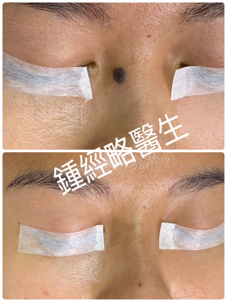
患者為一位 32 歲女性,無重大過去病史或家族病史。她因右側鼻樑斜坡出現黑色皮膚病變而求診,該病變最早於她 7 歲時發現。雖然病變在童年時期緩慢增長,但在過去十年間保持完全穩定。患者表示無出血、分泌物、潰瘍、發紅、腫脹、疼痛、壓痛或瘙癢。她要求切除純粹出於美觀考量。
體格檢查
檢查顯示,病變大小為 0.7 厘米 × 0.6 厘米,凸起高度約 0.2 厘米。病變呈對稱橢圓形,長軸呈垂直方向。邊緣清晰且明確。表面呈現正常的皮膚紋理與毛孔。顏色呈均勻的黑色。觸診時質地正常,無壓痛、疼痛或周圍紅斑。
初步診斷
良性混合痣(Benign compound nevus)。
手術方案與過程
在此區域切除皮膚病變面臨多項解剖學上的挑戰。我們使用 UltraPulse CO₂ 超脈衝二氧化碳激光進行精準雷射切除,以徹底清除位於根部的所有黑色素細胞巢。激光氣化過程無焦化且無活動性出血,確保了極高的黑色素細胞巢完整清除率。
然而,激光切除後留下了深度約 0.3 厘米的缺損。若在此深度進行二期癒合(自然癒合),必然會導致不合宜的凹陷性萎縮疤痕。因此,我們決定採用一期縫合修復傷口。
為減低縫合線張力並防止狗耳狀畸形(dog-ear deformities),我們將傷口以 3:1 的長寬比進行手術延長。隨後進行針對性的皮下剝離(undermining)。接著,使用 6-0 可吸收縫線縫合皮下組織。最後,使用 5-0 尼龍線進行皮膚對合縫合。
術後結果
術後 14 天順利拆除尼龍縫線,無任何併發症。在 6 個月的隨訪中,患者的傷口呈現出極其淡化、幾乎不可見的疤痕。
Case Presentation
This 32-year-old female has no significant past medical or family history. She presented with a black skin lesion on the right lateral slope of her nasal bridge, which first appeared when she was 7 years old. Although it grew slowly during childhood, it remained completely stable over the past decade. She noted no bleeding, discharge, ulceration, redness, swelling, pain, tenderness, or itchiness. She requested removal strictly for cosmetic reasons.
Physical Examination
On examination, the lesion measured 0.7 cm × 0.6 cm with a 0.2 cm elevation. It was symmetrical and oval, with its long axis oriented vertically. The border was clear and well-defined. The surface displayed normal skin markings and pores. Coloration was homogenously black. Palpation revealed a soft tissue texture with no tenderness, pain, or surrounding erythema.
Impression & Diagnosis
Benign compound nevus.
Surgical Approach & Procedure
Excising a lesion in this region presented several anatomical challenges. We performed precise laser excision using an UltraPulse CO₂ laser to clear all nested melanocytes at the root completely. The laser ablation produced zero charring and zero active bleeding, ensuring a exceptionally high rate of complete melanocyte removal.
However, laser removal left a deep defect measuring approximately 0.3 cm. Healing by secondary intention at this depth would inevitably cause an unsightly, depressed atrophic scar. Therefore, we elected to close the wound primarily.
To reduce suture line tension and prevent dog-ear deformities, we surgically extended the wound along a 3:1 length-to-width ratio. We then performed targeted subcutaneous undermining. Next, we closed the subcutaneous layer using absorbable 6-0 sutures. Finally, we re-approximated the skin edges using 5-0 nylon.
Outcome
We removed the nylon sutures 14 days post-operatively without complication. At the 6-month follow-up, the patient demonstrated an exceptionally subtle, almost invisible scar.
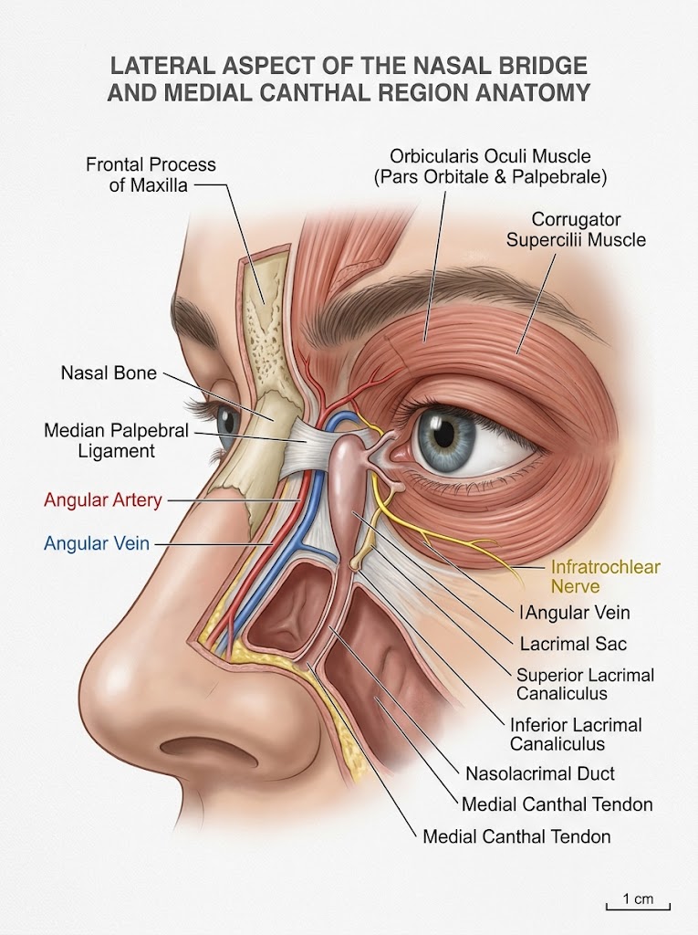
在鼻樑側翼(內眥/鼻旁斜坡)切除病變需要周詳的規劃,因為淺層及深層皮下組織中密集分佈著關鍵的血管網絡、運動/感覺神經以及具功能性的淚器結構。
該區域的關鍵解剖結構
1. 血管(高出血及栓塞風險)
- 角動脈與角靜脈(Angular Artery & Vein): 面動脈的終端分支,沿鼻側斜坡垂直行走,位於內眥內側約 6–8 毫米處。它部位於鼻肌及眼輪匝肌纖維之內或緊貼其深層。
- 眼動脈吻合網(Ophthalmic Artery Anastomoses): 靠近內眥處,角動脈直接與鼻背動脈及滑車上動脈(經眼動脈來自頸內動脈系統的分支)吻合。
- 手術重要性: 無意的損傷可導致劇烈的動脈出血或血腫形成。在進行醫美注射(如微整填補劑)或局部麻醉時,此處的血管內注射帶有逆行性栓塞至視網膜中央動脈的風險,從而導致失明。
2. 神經(感覺與運動)
- 滑車下神經(Infratrochlear Nerve,三叉神經 V1 分支): 行走於內眥上方內側,負責鼻樑上部、內側眼瞼及淚囊區域皮膚的感覺。
- 鼻外神經(External Nasal Nerve,三叉神經 V1 分支): 負責鼻下斜坡及鼻背皮膚的感覺。
- 眶下神經分支(Infraorbital Nerve branches,三叉神經 V2 分支): 遠端淺層末梢負責鼻下側斜坡皮膚的感覺。
- 面神經顴支/頰支(Facial Nerve Zygomatic/Buccal Motor Branches): 支配眼輪匝肌(以及提上唇鼻翼肌和鼻肌)。過深或過廣的切除可能導致局部面部肌肉無力或眼瞼不對稱。
3. 淚器(淚液引流系統)
- 淚囊與鼻淚管(Lacrimal Sac & Nasolacrimal Duct): 位於內眥韌帶後方的淚囊窩內。
- 上、下淚小管(Superior & Inferior Canaliculi): 沿上下眼瞼內側緣水平行走,通往淚囊。
- 手術重要性: 切口或深層剝離若過於向外側延伸至眼瞼韌帶,有割裂淚小管或淚囊的風險,從而引致慢性溢淚(淚水過多溢出)及淚囊炎。
4. 淋巴引流
- 內眥及鼻部淋巴管(Medial Canthal & Nasal Lymphatics): 此區域的淋巴管主要向下及向外側引流至下頜下淋巴結,較上方的通道則引流至耳前/腮腺淋巴結。
皮膚切除時的手術考量與警戒事項
| 風險領域 | 預防措施 / 手術技巧 |
| 剝離深度(Depth of Dissection) | 若病變純粹屬於表皮或皮內,應維持在肌肉層(眼輪匝肌/鼻肌)上方的皮下平面。除非因惡性腫瘤需要確保安全邊緣,否則應避免直接在骨膜上方進行深層銳性剝離。 |
| 止血(Hemostasis) | 由於角血管網絡豐富,預期會有即時出血。在內眥附近應使用細尖雙極電灼(bipolar cautery)而非單極電灼,以保護鄰近的神經分支及眼球。 |
| 切口方向(Incision Orientation) | 切口應平行於放鬆皮膚張力線(RSTLs),該線條通常沿鼻斜坡呈斜向或對角線走向。垂直疤痕可能導致蹼狀攣縮(內眥贅皮褶皺),或將內眥向內/向下拉扯。 |
| 淚器保護(Lacrimal Protection) | 辨識並保留內眥韌帶。在未確認淚囊位置前,切勿盲目切割內眥深層的纖維組織。 |
| 眼瞼外翻與牽引(Ectropion & Eyelid Traction) | 避免對下眼瞼產生過度傷口張力,否則可導致牽引性眼瞼外翻。相較於張力下的直接縫合,通常首選皮瓣修復(如雙葉皮瓣、菱形皮瓣或移位皮瓣)或全層植皮。 |
編者按:
鍾經略醫生之英國臨床皮膚科碩士及英國美容醫學碩士課程案例針對鼻旁斜坡(Nasal Sidewall)及內眥(Medial Canthus)區域的良性色素痣或惡性皮膚腫瘤,無論採用激光治療或外科手術切除,均具高度臨床挑戰性,術前評估與操作必須極為審慎。由於該解剖區域的組織結構精細且血管神經密集,容易導致以下潛在併發症:
1. 血管損傷與組織缺血會引發局部缺血甚至皮膚及軟組織壞死,不僅影響外觀,嚴重者更可能波及周邊面部器官的正常功能。
2. 神經功能受損會導致局部皮膚感覺麻木、缺失,甚至引發局部面部肌肉無力或雙側眼瞼不對稱。
3. 淚道系統受損會誘發慢性溢淚及淚囊炎等長期後遺症。
鑑於上述解剖複雜性,此部位的病變脫除絕非單純的基礎處置。為確保療效並將手術風險降至最低,建議患者在選擇主診醫生時應審慎評估其專業資歷——理想人選應具備深厚的臨床皮膚病學訓練背景(如相關碩士或以上學歷,並擁有多年臨床皮膚外科經驗)及嚴謹的美容醫學專業資格(碩士或以上學歷),以提供最安全且具針對性的治療方案。
Excising a lesion at the lateral aspect of the nasal bridge (the medial canthal/paranasal slope) requires careful planning due to a dense concentration of critical vascular networks, motor/sensory nerves, and functional lacrimal apparatus structures running through the superficial and deep subcutaneous layers.
Key Anatomical Structures in the Region
1. Blood Vessels (High Risk of Bleeding & Embolization)
- Angular Artery & Vein: The terminal branch of the facial artery runs vertically along the lateral slope of the nose, approximately 6–8 mm medial to the inner canthus. It lies within or just deep to the nasalis and orbicularis oculi muscle fibers.
- Ophthalmic Artery Anastomoses: Near the medial canthus, the angular artery anastomoses directly with the dorsal nasal artery and supratrochlear artery (branches of the internal carotid system via the ophthalmic artery).Surgical Significance: Inadvertent trauma can cause brisk arterial bleeding or hematoma formation. In cosmetic filler or local anesthetic procedures, intravascular injection here carries a risk of retrograde embolization to the central retinal artery leading to blindness.
2. Nerves (Sensory & Motor)
- Infratrochlear Nerve (CN V1 branch): Runs just superior-medial to the inner canthus, supplying cutaneous sensation to the skin of the upper nose bridge, medial eyelids, and lacrimal sac region.
- External Nasal Nerve (CN V1 branch): Supplies skin over the lower slope and dorsum of the nose.
- Infraorbital Nerve branches (CN V2): Distal superficial twigs supply skin along the inferior-lateral slope of the nose.
- Facial Nerve Zygomatic/Buccal Motor Branches: Innervate the orbicularis oculi (levator labii superioris alaeque nasi and nasalis muscles). Deep or wide excision can cause localized facial muscular weakness or eyelid asymmetry.
3. Lacrimal Apparatus (Tear Drainage)
- Lacrimal Sac & Nasolacrimal Duct: Housed within the lacrimal fossa behind the medial palpebral ligament.
- Superior & Inferior Canaliculi: Run horizontally within the medial margins of the upper and lower eyelids toward the lacrimal sac.Surgical Significance: Incisions or deep dissections carried too far laterally toward the palpebral ligament risk lacerating the lacrimal canaliculi or sac, leading to chronic epiphora (overflow of tears) and dacryocystitis.
4. Lymphatic Drainage
- Medial Canthal & Nasal Lymphatics: Lymphatic vessels in this zone drain primarily downward and laterally toward the submandibular lymph nodes, with superior channels draining into the preauricular/parotid lymph nodes.
Surgical Considerations & Red Flags During Skin Excision
| Risk Area | Precaution / Surgical Technique |
| Depth of Dissection | Stay in the subcutaneous plane above the muscular layer (orbicularis oculi / nasalis) if the lesion is purely epidermal or intradermal. Avoid deep sharp dissection directly over the periosteum unless margin clearance for malignancy requires it. |
| Hemostasis | Expect prompt bleeding due to the angular vessel network. Use fine-tip bipolar cautery near the inner canthus rather than monopolar to protect nearby nerve branches and the eye. |
| Incision Orientation | Orient incisions parallel to Relaxed Skin Tension Lines (RSTLs), which generally run obliquely or diagonally along the nasal slope. Vertical scars can cause webbed contractures (epicanthal folding) or pull the inner canthus medially/inferiorly. |
| Lacrimal Protection | Identify and preserve the medial palpebral ligament. Do not blindly divide fibrous tissue deep to the medial canthus without identifying the lacrimal sac location. |
| Ectropion & Eyelid Traction | Avoid excessive wound tension toward the lower eyelid, which can cause traction ectropion. Flap closure (e.g., bilobed, rhomboid, or transposition flap) or full-thickness skin grafting is often preferred over primary closure under tension. |
Editor’s Note:
Removing benign moles or skin cancers on the side of the nose (nasal sidewall) and inner corner of the eye (medial canthus) is challenging. Whether a doctor uses a laser or surgery, the procedure requires careful planning and precision. Furthermore, this area contains delicate tissues packed with blood vessels and nerves. As a result, treatments carry several serious risks:
- Blood Vessel Damage can cause skin and tissue death (necrosis), affecting both appearance and, in severe cases, the function of nearby facial features.
- Nerve Damage can lead to skin numbness, partial facial muscle weakness, or uneven eyelids.
- Tear Duct Damage can cause long-term issues like chronic watery eyes and tear sac infections.
Therefore, removing lesions here is far from a simple routine procedure. To get the best results and minimize risks, you should carefully check your doctor’s qualifications. Specifically, look for a specialist with extensive training in clinical dermatology (a Master’s degree or higher, plus over 10 years of experience). They should also hold formal credentials in aesthetic medicine (a Master’s degree or higher). Ultimately, choosing a qualified expert ensures a safe, tailored treatment plan.