
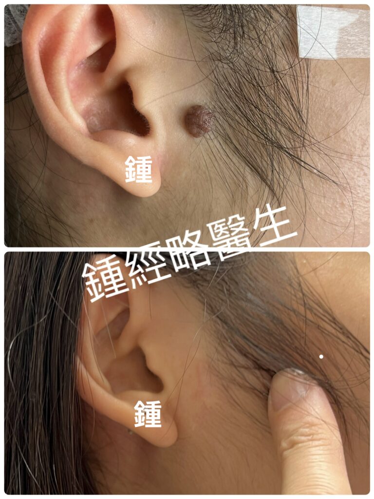
耳前區脱痣 :耳前部位進行切痣手術時,解剖構造相當複雜且精細。由於該區域緊鄰顏面神經分支、重要血管以及腮腺,血管與神經破壞確實是關鍵的考量之一,但透過專業的解剖層次控管與手術設計,這些風險是可以被有效控制的。
1. 血管與神經破壞的風險評估
耳前區的切痣手術是否會傷害到神經或血管,主要取決於痣的深度、大小以及切除時的解剖層次:
顏面神經(Facial Nerve)
- 涉及分支:耳前區下方有顏面神經主幹從莖乳孔出頭後進入腮腺,並分出顳支(Temporal branch)與顴骨支(Zygomatic branch)向前方及上方走行。
- 損傷風險與後果:若傷及顏面神經分支,可能導致抬眉困難、眼瞼無法完全閉合或嘴角下垂/麻痺。
- 安全防線:顏面神經的分支在耳前區大多走行於深筋膜(SMAS層)下方或腮腺組織內。一般的皮膚痣(良性痣或常見色素痣)通常僅涉及表皮層與真皮層,切除時若嚴格維持在皮下脂肪淺層進行,極少會傷及顏面神經。
耳大神經與耳顳神經(Sensory Nerves)
- 耳顳神經(Auriculotemporal Nerve)與耳大神經前支(Great Auricular Nerve)負責耳前及顳區的感覺。
- 損傷後果:若切除較深,可能造成耳前或局部皮膚暫時性或永久性的麻木感、知覺異常。
血管(Blood Vessels)
- 涉及血管:淺顳動靜脈(Superficial Temporal Vessels)走行於耳前前方。
- 損傷風險:淺層切除若碰到微血管出血,可透過電燒(Electrocautery)快速止血;若切得較深碰到淺顳動靜脈,出血量會較多,但經驗豐富的醫師能清晰辨識並進行結紮或電燒止血,通常不會造成嚴重的系統性後果。
2. 耳前區脱痣的其他核心考量
除神經與血管外,耳前區切痣還需要考量以下幾個臨床要素:
| 考量項目 | 說明與臨床處理 |
| 切口方向與皮紋(Langer’s Lines) | 顏面的皮膚張力線在耳前區大多呈垂直方向。切口應順著耳前的自然褶皺(如耳屏前緣)設計,縫合後疤痕能最大程度隱藏。 |
| 疤痕與張力(Scarring & Tension) | 耳前皮膚張力適中,但若痣較大,直接縫合可能拉扯到耳屏(Tragus)導致耳部變形。較大的痣可能需要局部皮瓣移植(Local Flap)或分階段切除。 |
| 耳軟骨與腮腺(Cartilage & Parotid Gland) | 痣若較深,需避免傷及深層的耳軟骨膜(否則易引發發炎或變形)及腮腺包膜(避免腮腺瘻管或唾液積聚)。 |
| 病理組織檢查(Pathology) | 任何切除的色素痣均應送交病理切片檢查,以排除基底細胞癌、黑色素瘤等惡性病變的可能。 |
3. 臨床建議
- 評估與執行醫師:耳前區構造精細,建議尋求具有皮膚科碩士及醫學美容碩士(或以上)雙重資格的醫師進行評估與手術。
- 術前評估:醫師會在術前觸診並評估痣的邊界與深度。對於純表淺的痣,亦可由醫師評估是否適合採用雷射(如雷射無法完全清除或懷疑惡性時,仍以手術切除為主)。
- 術後照護:術後遵照醫囑保持傷口清潔乾燥,按時換藥與拆線,並在拆線後配合抗疤產品(如矽膠凝膠或矽膠貼片)以達最佳修復效果。
Moles Removal in the Preauricular Region
Removing a mole from the preauricular region (the area in front of the ear) requires precise technique because the underlying anatomy is intricate. This region sits close to facial nerve branches, major blood vessels, and the parotid gland. As a result, preventing nerve and vascular damage is a primary concern. Nevertheless, experienced surgeons can control these risks effectively through precise anatomical depth control and proper surgical planning.
1. Risk Assessment: Nerve and Vascular Damage
Whether a preauricular mole removal injures nerves or blood vessels depends on three factors: the mole’s depth, its size, and the surgical plane chosen during excision.
Facial Nerve
- Involved Branches: The main trunk of the facial nerve emerges from the stylomastoid foramen, enters the parotid gland, and gives off the temporal and zygomatic branches. These branches run anteriorly and superiorly across the preauricular region.
- Risks and Consequences: Damaging these facial nerve branches can cause drooping eyebrows, incomplete eyelid closure, or corner-of-the-mouth asymmetry and weakness.
- Safety Buffer: In this area, facial nerve branches mostly run beneath the deep fascia (SMAS layer) or within the parotid gland. Common skin moles (benign nevi) typically involve only the epidermis and dermis. Therefore, by staying in the superficial subcutaneous fat layer, surgeons rarely compromise the facial nerve.
Sensory Nerves (Auriculotemporal and Great Auricular Nerves)
- Involved Branches: The auriculotemporal nerve and the anterior branch of the great auricular nerve supply sensation to the preauricular and temporal regions.
- Risks and Consequences: Deeper excisions can injure these sensory fibers, which leads to temporary or permanent numbness and paresthesia in the local skin.
Blood Vessels
- Involved Vessels: The superficial temporal artery and vein run directly in front of the ear.
- Risks and Consequences: Superficial cuts may hit small capillaries, which surgeons easily stop with electrocautery. Deeper cuts might encounter the main superficial temporal vessels and cause heavier bleeding. However, skilled surgeons readily identify these vessels and secure them using ligation or electrocautery, preventing serious systemic complications.
2. Additional Clinical Considerations
Beyond nerves and blood vessels, surgeons evaluate several critical factors before operating on this region:
| Clinical Factor | Explanation and Management |
| Incision Line & Tension Lines | Facial tension lines (Langer’s lines) run mostly vertically in front of the ear. Surgeons align incisions with natural creases, such as the pretragal fold, to hide the final scar effectively. |
| Scarring & Tension Control | Although preauricular skin tension is moderate, excising large moles can pull on the tragus and deform the ear structure. Consequently, surgeons may use local skin flaps or staged excision for larger lesions. |
| Ear Cartilage & Parotid Gland | Deep excisions must avoid the underlying perichondrium to prevent inflammation or deformity. Additionally, surgeons must preserve the parotid capsule to prevent salivary fistulas or fluid collections. |
| Pathology Examination | Surgeons send every excised mole for histological examination. This routine check rules out malignancies like basal cell carcinoma or melanoma. |
3. Clinical Guidance
- Choose the Right Specialist: Because preauricular anatomy is complex, consult a plastic surgeon or dermatologic surgeon for evaluation and treatment.
- Preoperative Evaluation: Before operating, the surgeon will palpate the site to evaluate the mole’s borders and depth. For purely superficial moles, laser treatment may be an option. However, if the mole sits deep or shows suspicious features, surgical excision remains the standard of care.
- Postoperative Care: Keep the wound clean and dry according to instructions. Attend all follow-up visits for wound care and suture removal. Afterward, use anti-scar treatments—such as silicone gel or silicone sheets—to ensure optimal aesthetic healing.
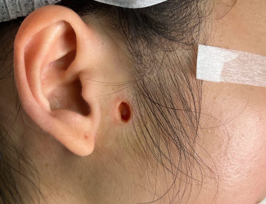
上圖顯示經二氧化碳雷射切除良性痣後之創面。雷射切割範圍與病變大小完全吻合,未過度切除正常組織。創緣熱傷害極小,周邊組織活力良好。傷口完全止血且無焦碳形成。目視確認深層黑色素細胞巢已完整清除、無殘留,復發率極低。創底組織結構正常,邊緣與基底組織修復能力良好。
The photo above shows the wound bed following CO2 laser excision of a benign nevus. The laser resection margin completely corresponds to the lesion size without excessive removal of normal tissue. Thermal damage to the wound edges is minimal, preserving strong tissue viability in the surrounding area. Complete hemostasis was achieved with no char formation. Visual inspection confirms complete clearance of deep melanocytic nests with no residual tissue, yielding a very low recurrence rate. The wound base exhibits normal tissue architecture, with high regenerative potential of both the marginal and basal tissues.
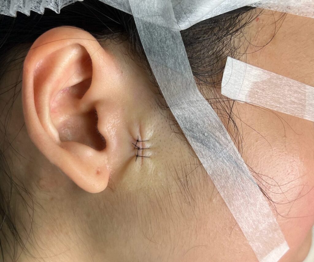
上圖為傷口閉合後之創面。深部採用 6-0 可吸收縫線進行皮下層縫合,表皮則採用 5-0 尼龍縫線進行三針單純間斷縫合。
The photo above demonstrates the wound bed after closure. Deep subcutaneous closure was performed using 6-0 absorbable sutures, and the outer skin layer was approximated with three simple interrupted 5-0 nylon sutures.
個案彙報
患者資料: 33歲女性
既往病史: 無特殊紀錄
家族病史: 無特殊紀錄
現病史
患者主訴耳屏前(左/右耳耳屏前方)有一處歷時已久的緩慢增長皮膚病變。該病變最早於約5歲時被發現。在過去十年間,其狀況維持高度穩定,顏色、色澤深淺、形狀、邊界及大小均無突發性改變。患者無疼痛、壓痛、瘙癢、紅斑、腫脹或潰瘍病史。是次主要因美容考量要求進行手術切除。
身體檢查
- 位置: 耳屏前區域(耳屏前方)
- 尺寸: 0.5 cm × 0.4 cm,高出皮膚表面 0.3 cm
- 形態: 呈橢圓形、對稱、均勻淺棕色之丘疹,邊界清晰且規則
- 表面特徵: 皮膚表面完整且無潰瘍跡象;病變處有 6 至 10 條細小終毛生長
- 觸診: 無壓痛,周圍無硬化或發炎現象
臨床診斷
良性混合型痣伴終毛增生
手術過程與處理
- 手術名稱: 超脈衝二氧化碳雷射磨皮術配合極小張力一期縫合
- 技術與視覺輔助: 手術全程於 3.5 倍手術放大鏡輔助下進行精準熱氣化磨皮,並配備醫用級吸煙器。氣化深度達真皮深層,直至徹底清除所有手術放大鏡可見之黑色素細胞巢(痣根),以降低復發風險。
- 術中情況: 止血效果極佳,全程無焦炭形成或術中出血。未造成任何血管或面神經分支損傷。
- 傷口縫合: 完整氣化黑色素細胞巢後,將局部皮下組織進行輕微游離。傷口採分層縫合:皮下層使用 6-0 可吸收縫線對合,表皮則以 5-0 尼龍線進行 3 針間斷縫合。
術後狀況與預後
- 拆線: 術後 2 週順利拆除間斷尼龍縫線。
- 癒合: 傷口癒合完整且無併發症。
- 術後跟進(6個月): 術後 6 個月臨床覆診顯示,肉眼觀察下疤痕極小且幾乎不可見,達到優異之美觀復原效果,且無臨床復發跡象。
Case Presentation
Patient Identification: 33-year-old female
Past Medical History: Unremarkable
Family History: Unremarkable
History of Present Illness
The patient presented with a long-standing, slowly growing preauricular skin lesion situated anterior to the tragus of the left/right ear. The lesion was first noticed around age 5. Over the past decade, it had remained completely stable with no sudden changes in color, shade, shape, border, or size. The patient reported no history of pain, tenderness, pruritus, erythema, swelling, or ulceration. She requested surgical excision primarily for cosmetic concerns.
Physical Examination
- Location: Preauricular region (anterior to the tragus)
- Dimensions: 0.5 cm×0.4 cm, elevated 0.3 cm above skin level
- Morphology: Oval, symmetrical, homogenous light brown papule with well-defined, regular borders
- Surface Characteristics: Intact skin surface with no evidence of ulceration; 6–10 fine terminal hairs projecting from the lesion
- Palpation: Non-tender, no surrounding induration or inflammation
Clinical Impression
Benign compound nevus with terminal hypertrichosis.
Surgical Procedure & Management
- Procedure: Ultrapulse CO2 laser ablation and minimal-tension primary closure
- Technique & Visualization: Precise thermal ablation was executed under 3.5× magnification loupe assistance, paired with a medical-grade smoke evacuator. The lesion was ablated down to the deep dermal layers until all macroscopically evident (under 3.5x magnification) melanocytic nests (mole roots) were fully cleared to minimize recurrence risk.
- Intraoperative Findings: Excellent hemostasis with zero wound charring or intraoperative bleeding. No vascular or facial nerve branch injury occurred.
- Closure: Following complete ablation of melanocytic nests, the local subcutaneous tissue was lightly undermined. Closure was performed in layers: subcutis approximated using 6-0 absorbable sutures, followed by epidermal adaptation using three interrupted 5-0 nylon sutures.
Postoperative Course & Outcome
- Removal of Sutures: Interrupted nylon stitches were removed at 2 weeks postoperatively.
- Healing: Wound healing was complete and uneventful.
- Follow-up (6 Months): Clinical examination at 6 months post-procedure demonstrated exceptional aesthetic recovery with a minimal, virtually imperceptible scar under normal visual inspection. No clinical signs of recurrence were observed.