
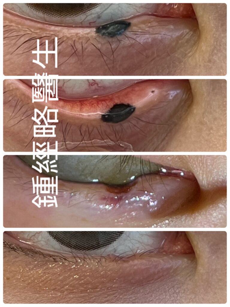
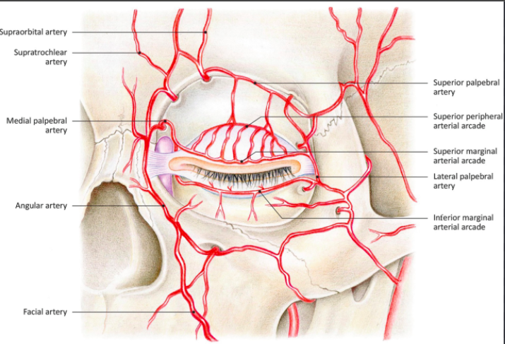
The anatomical position of inferior marginal palpebral arch 下眼皮邊緣動脈拱(或稱下瞼邊緣動脈弓)的位置
下眼瞼緣墨痣進行 CO₂ 激光氣化(下眼皮睫毛邊緣脫痣)時保護邊緣動脈拱(Marginal Arterial Arcade)之策略
前言
清除眼瞼緣的良性墨痣,必須在「徹底去痣」、「保留結構」與「保護眼瞼功能」三者之間取得精確平衡。
下眼瞼緣是一個結構非常緊湊且複雜的部位。在不到兩毫米的狹窄範圍內,集結了毛囊、瞼板組織、油腺(瞼板腺)出口,以及極為關鍵的微血管網絡。
其中,邊緣動脈拱(Marginal arterial arcade) 扮演著眼瞼緣的生命線。這條血管負責為眼瞼肌肉、腺體網絡以及黏膜交界處提供養分。因此,當醫生使用 CO₂ 激光等高精準度儀器(如超脈衝 Ultrapulse 模式)時,若過多熱力滲透,便有機會破壞這條動脈拱。熱傷害一旦發生,可導致局部組織壞死、眼瞼緣缺口畸形、疤痕性眼瞼外翻,或眼油腺萎縮。
歸根究底,要做到既能安全進行下眼皮睫毛邊緣脫痣,又能保護這條動脈拱,關鍵在於三個步驟:掌握血管深度、精確控制熱能,以及運用靈巧的手術技巧。
1. 邊緣動脈拱的解剖位置與深度
首先,醫生必須清楚掌握這條動脈拱相對於眼瞼標誌的三維位置與深度。
- 血管來源: 下眼瞼外側動脈與內側動脈在眼瞼緣附近會合,共同組成了這條下眼瞼邊緣動脈拱。
- 行走路線: 這條動脈拱橫向穿過下眼瞼,與眼瞼邊緣平行。具體而言,它位於眼瞼邊緣下方約 1.0 至 1.5 毫米 處。
- 所在層次與深度: 此外,動脈拱位於瞼板前方的疏鬆組織層中,同時藏於眼輪匝肌(特別是 Riolan 氏肌)的深層。整體而言,視乎患者年齡及皮膚鬆弛程度,它位於皮膚表面下方約 1.0 至 2.0 毫米 深之處。
2. 激光熱傷害的病理機制
CO₂ 激光發射波長為 10,600 納米(nm)的光束。由於細胞內外的水分會迅速吸收這種波長,目標組織會瞬時沸騰、破裂並氣化。
然而,若果多餘的熱力透過墨痣滲透至深層的動脈壁,便會引發以下三種破壞性反應:
- 血管內凝血與血栓: 首先,直接的熱力會損害血管內皮細胞及紅血球。結果會形成血栓,阻斷局部血液循環。
- 膠原蛋白收縮與管腔閉塞: 其次,高溫會使血管壁的膠原蛋白變性。結果導致血管壁塌陷並永久封閉。
- 缺血性組織壞死: 最後,缺乏血液供應會使附近的瞼板膠原及油腺缺氧衰竭。因此,組織隨後會壞死脫落,造成眼瞼缺口及不規則疤痕。
3. 保護邊緣動脈拱的手術策略
要在 CO₂ 激光氣化過程中保護這條動脈拱,醫生必須結合適當的參數設定、主動冷卻、動態組織拉開以及持續的深度監測。
A. 參數優化:極短脈衝與熱釋放時間
重點在於限制熱力擴散,使其低於周圍真皮層的「熱釋放時間(TRT)」(約 0.1 至 1.0 毫秒)。
- 超脈衝模式(Ultrapulse / Superpulse): 首先,在眼瞼緣附近切忌使用連續波(CW)模式,因為連續波會導致熱力無法控制地擴散。相反,應使用超脈衝模式,在極短的時間內(小於 1 毫秒)釋放高峰值功率。這樣,激光可以在熱力傳導至深層動脈拱之前,便已將目標組織氣化。
- 能量密度與光斑大小: 此外,應選擇較細的光斑(0.1 至 0.2 毫米)以精準鎖定墨痣。當氣化接近深層瞼板邊界時,適當降低總能量(2.0 至 5.0 J/cm²)。
B. 熱能管理與主動冷卻
此外,醫生必須主動冷卻周圍組織,以阻斷熱力傳導:
- 冰生理鹽水保護: 首先,在手術期間不斷使用冰凍生理鹽水沖洗周圍區域。這種冷液體能在熱能滲入深層動脈拱之前,將多餘熱力帶走。
- 眼球防護: 術前放入金屬角膜保護罩(corneal shield)。這樣既能保護眼球,又能提供穩固的支撐,方便進行精細的手術定位。
C. 機械性拉開與水分離技術(Hydrodissection)
此外,醫生可以透過手動操作,拉大墨痣底部與下方動脈之間的安全距離:
- 外翻與對向拉張: 首先,使用無齒精細鉗或皮膚鉤,輕柔地將眼瞼緣向外翻離眼球。這個拉伸動作能將表淺的墨痣拉離深層血管網,從而增加解剖上的安全距離。
- 水分離技術(Hydrodissection): 其次,將局部麻醉藥(例如含有 1:100,000 腎上腺素的 2% 利多卡因)直接注射至墨痣下方的瞼板前空間。這層液體墊能將皮膚與深層動脈拱之間的距離增寬數百微米,發揮阻隔激光熱力的作用。
D. 逐層氣化與停點判定
最後,切忌一次過進行深層氣化,而應在高倍放大鏡(3.5倍 至 5.0倍)或手術顯微鏡下分步進行:
- 表層削平(Debulking): 首先氣化墨痣凸出的部分,使其與周圍皮膚平齊。
- 清除碳化殘渣: 每完成一次激光照射後,必須用濕潤的鹽水棉花輕輕擦走黑色碳化殘渣。清除殘渣至關重要,因為殘留的碳化物會積聚熱力,像烙鐵一樣將高溫傳導至深層組織。
- 深層停點判定: 當氣化接近真皮深層時,將激光切換至單脈衝模式。一旦看到乾淨、有光澤、無色素的瞼板前筋膜出現,便需立即停止,切勿穿透至眼輪匝肌層。
4. 術後護理與預後
當邊緣動脈拱得到妥善保護,完好的微循環能促進傷口快速平滑地癒合:
- 快速表皮上皮化: 由於局部血流維持正常,健康的表皮細胞能從鄰近皮膚迅速遷移,在 7 至 14 天內完成傷口覆蓋。
- 最小化疤痕收縮: 此外,維持血液供應可防止缺血性纖維化。因此,患者不會出現眼瞼缺口、睫毛脫落(Madarosis)或眼瞼外翻等結構性問題。
- 術後護理: 最後,每天塗抹眼用抗生素軟膏。軟膏能保持傷口濕潤、消退發炎,並在眨眼時減少摩擦。
結論
總結而言,使用 CO₂ 激光進行下眼皮睫毛邊緣脫痣手術,需要極高的精準度與清晰的解剖概念。邊緣動脈拱是眼瞼邊緣的生命線。只要善用超脈衝模式、水分離技術、冰鹽水冷卻以及清晰的逐層氣化技巧,醫生便能在完全保護眼瞼結構與功能的前提下,徹底去痣。
Strategic Preservation of the Marginal Arterial Arcade During CO2 Laser Vaporization of Lower Eyelid Margin Nevi
Introduction
Ablating a benign skin nevus on the palpebral margin requires a careful balance. The surgeon must clear the lesion completely, preserve structural integrity, and protect lid function.
The lower eyelid margin is a compact, complex structure. Within less than two millimeters, hair follicles, tarsal plate connective tissue, Meibomian gland orifices, and critical microvessels all converge.
Consequently, the marginal arterial arcade maintains the vascular lifeline of the lid margin. This vessel feeds the pretarsal muscles, glandular network, and mucocutaneous junction. Therefore, when surgeons use high-precision tools like CO2 lasers in Ultrapulse or superpulsed mode, excess heat can destroy this arcade. As a result, thermal injury causes local necrosis, notch deformities, cicatricial ectropion, or Meibomian gland loss.
Ultimately, protecting the marginal arcade while eliminating a benign nevus requires three key steps: understanding vascular depth, controlling thermal energy, and executing precise manual techniques.
1. Topographical & Vascular Anatomy of the Marginal Arcade
First, surgeons must visualize the three-dimensional depth and trajectory of the marginal arcade relative to key lid landmarks.
- Vascular Origins: The inferior lateral palpebral artery (from the lacrimal system) and inferior medial palpebral artery (from the ophthalmic system) join together. As a result, this junction forms the lower eyelid marginal arcade.
- Anatomical Course: Next, the arcade runs horizontally across the lower lid, parallel to the edge. Specifically, it sits about 1.0 to 1.5 mm below the palpebral margin.
- Tissue Plane & Depth: Furthermore, the arcade sits in loose connective tissue directly in front of the lower tarsal plate. Simultaneously, it lies deep to the orbicularis oculi muscle fibers (near Riolan’s muscle). Overall, it lies 1.0 to 2.0 mm beneath the skin surface, depending on patient age and tissue laxity.
2. Pathophysiology of Laser Thermal Damage
The CO2 laser emits light at 10,600 nm. Because intracellular and extracellular water absorb this wavelength rapidly, the target tissue boils, bursts, and vaporizes.
However, if excess heat leaks past the nevus and reaches the arterial wall, three destructive events occur:
- Intravascular Coagulation & Thrombosis: First, direct heat injures endothelial cells and red blood cells. Consequently, blood clots form, blocking local blood flow.
- Collagen Contraction & Lumen Collapse: Next, heat denatures structural collagen within the vessel wall. As a result, the vessel wall collapses and closes permanently.
- Ischemic Tissue Necrosis: Finally, lack of blood supply starves nearby tarsal collagen and Meibomian glands. Therefore, tissue sloughs off later, creating lid notches and irregular scars.
3. Surgical Strategies for Vascular Preservation
To protect the marginal arcade during CO2 laser ablation, surgeons must combine smart power settings, active cooling, dynamic tissue retraction, and continuous depth monitoring.
A. Parameter Optimization: Short Pulses & Thermal Relaxation
Mainly, surgeons must limit heat diffusion so it stays below the thermal relaxation time (TRT) of adjacent dermis (0.1 to 1.0 ms).
- Ultrapulse / Superpulse Mode: First, avoid continuous wave (CW) mode near the lid edge, because CW mode causes uncontrolled heat spread. Instead, use Ultrapulse mode to deliver high peak power in ultra-short bursts (under 1 ms). Thus, the laser vaporizes target tissue faster than heat can diffuse into deeper planes.
- Energy Density & Spot Size: In addition, select a narrow spot size (0.1 to 0.2 mm) to isolate the nevus. Then, drop the total energy (2.0 to 5.0 J/cm2) as ablation approaches the deeper tarsal boundary.
B. Thermal Management & Active Cooling
In addition, surgeons must actively cool surrounding tissues to stop heat transfer:
- Chilled Saline Protection: First, flush the surrounding area continuously with ice-cold normal saline. This cold liquid absorbs excess heat before energy penetrates the deep arcade.
- Globe Protection: Meanwhile, place a corneal shield into the inferior fornix. Consequently, this shield protects the eye while providing a firm backing to support tissue positioning.
C. Mechanical Traction & Hydrodissection
Furthermore, surgeons can manually widen the safety gap between the nevus base and the underlying artery:
- Eversion & Counter-Traction: First, roll or evert the lid margin away from the globe using fine toothless forceps or a skin hook. This pulling motion stretches the tissue, moving the superficial nevus further away from the deep blood vessels.
- Hydrodissection: Next, inject local anesthetic (2% lidocaine with 1:100,000 epinephrine) directly under the lesion into the pretarsal space. Consequently, this fluid cushion expands the gap between the skin and the deep arcade by several hundred micrometers, insulating the artery from laser heat.
D. Layer-by-Layer Vaporization & Endpoints
Finally, instead of ablating deeply all at once, work step-by-step using high-magnification loupes (3.5times to 5.0times) or an operating microscope:
- Superficial Debulking: First, vaporize the raised portion of the nevus to level the surface.
- Debris Clearance: Next, wipe away carbonized char with a moist saline sponge after every pass. Removing char is crucial because leftover carbon traps heat and acts as a hot iron against deeper tissues.
- Deep Endpoint Verification: Then, switch to single-pulse mode as ablation nears the deep dermis. Stop immediately when clean, glistening, non-pigmented pretarsal fascia appears, before entering the orbicularis muscle layer.
4. Postoperative Care & Outcomes
When surgeons preserve the marginal arcade, intact microcirculation drives rapid, smooth healing:
- Fast Epithelialization: Because local blood flow remains strong, healthy epithelial cells migrate rapidly from adjacent skin, closing the wound within 7 to 14 days.
- Minimal Scarring: In addition, maintaining blood supply prevents ischemic fibrosis. Thus, patients avoid structural deformities like lid notches, eyelash loss (madarosis), or ectropion.
- Postoperative Protocol: Finally, apply a topical antibiotic ophthalmic ointment daily. This ointment keeps the wound moist, calms inflammation, and cushions the margin against friction from blinking.
Conclusion
In summary, ablating a lower eyelid margin nevus with a CO2 laser requires strict control and anatomical awareness. The marginal arterial arcade acts as the vital lifeline of the palpebral edge. Therefore, by using Ultrapulse mode, hydrodissection, active saline cooling, and deliberate layer-by-layer ablation, clinicians can eradicate benign nevi completely while fully preserving eyelid structure and function.