
【注意:本文章及真實病人照片均源自鍾經略醫生的臨床皮膚科碩士及美容醫學碩士課程案例,屬於原創作品。因此,本院嚴格保留所有版權。同時,我們禁止任何形式的轉載與複製。】
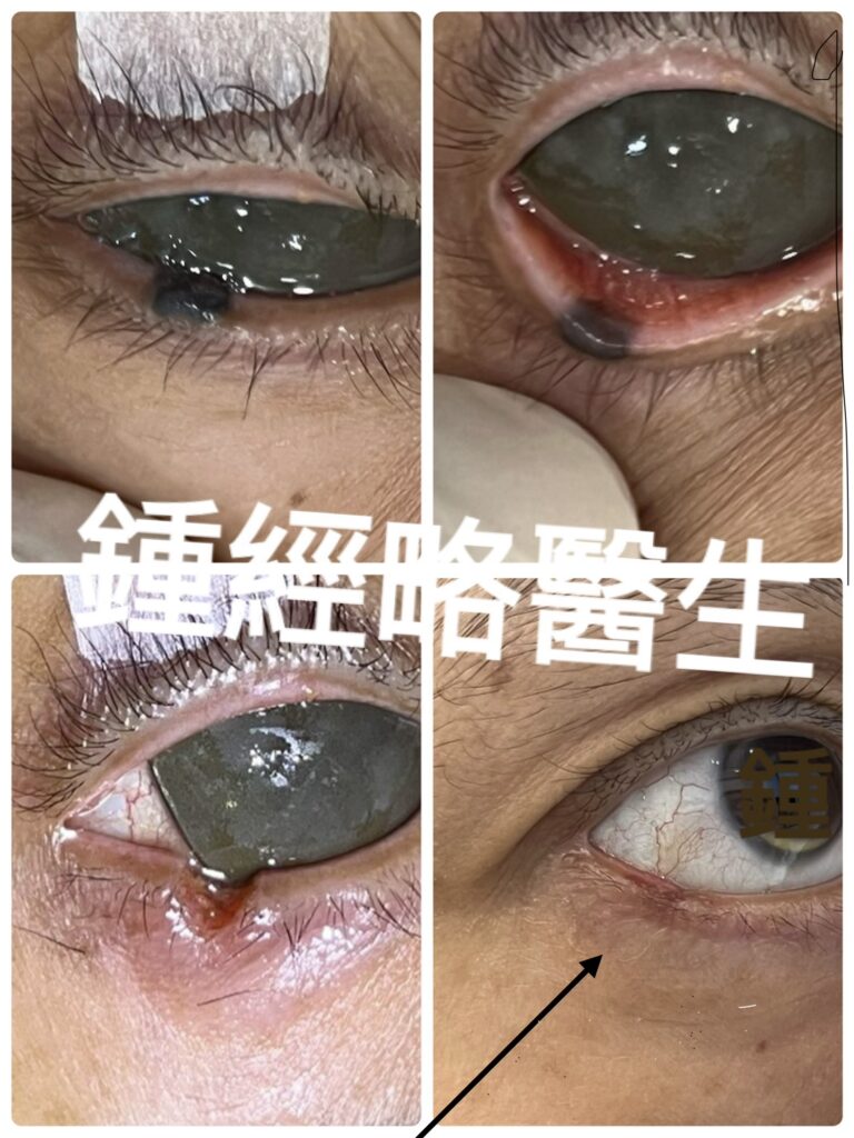
Operative Notes:
Patient: 53‑year‑old male
Procedure: CO₂ Laser Ablation of Lower Eyelid Mole with Post‑Laser Suturing
Surgeon: Dr Chung King Lueh MBBS (HK), MSc Aesthetic Medicine (QMUL), MSc Clin Derm (Lond)
Assistant: Joanne
Anesthesia: Local infiltration with 2% lidocaine with epinephrine
Chief Complaint
Unsightly skin mole on the right lower eyelid margin
History of Present Illness
The patient reports a pigmented lesion on the right lower eyelid margin that has been present for over 40 years. The lesion has grown slowly without sudden changes in morphology. He denies pain, irritation, discharge, or other associated symptoms. His primary concern is cosmetic appearance, with additional motivation for removal based on personal superstitious beliefs.
Past Medical History
- No significant past medical history
- No significant family history
Examination
- Lesion description: This homogeneous black skin lump measures approximately 0.4 cm × 0.2 cm. Furthermore, its longest axis runs parallel to the eyelid margin.
- Elevation: 0.1 cm above skin level.
- Extent: Spanning from the lower eyelid skin into the eyelid margin and extending down into the palpebral conjunctiva.
- Clinical signs: No erythema, ulceration, tenderness, or secondary changes noted.
- Systemic exam: Unremarkable.
Impression
Benign‑appearing pigmented lesion of the right lower eyelid margin, longstanding and asymptomatic, with no suspicious clinical features.
Plan
Excision/ laser removal of the lesion for cosmetic and personal reasons.
Histopathological examination not needed due to its benign nature.
Post‑operative wound care instructions to be provided.
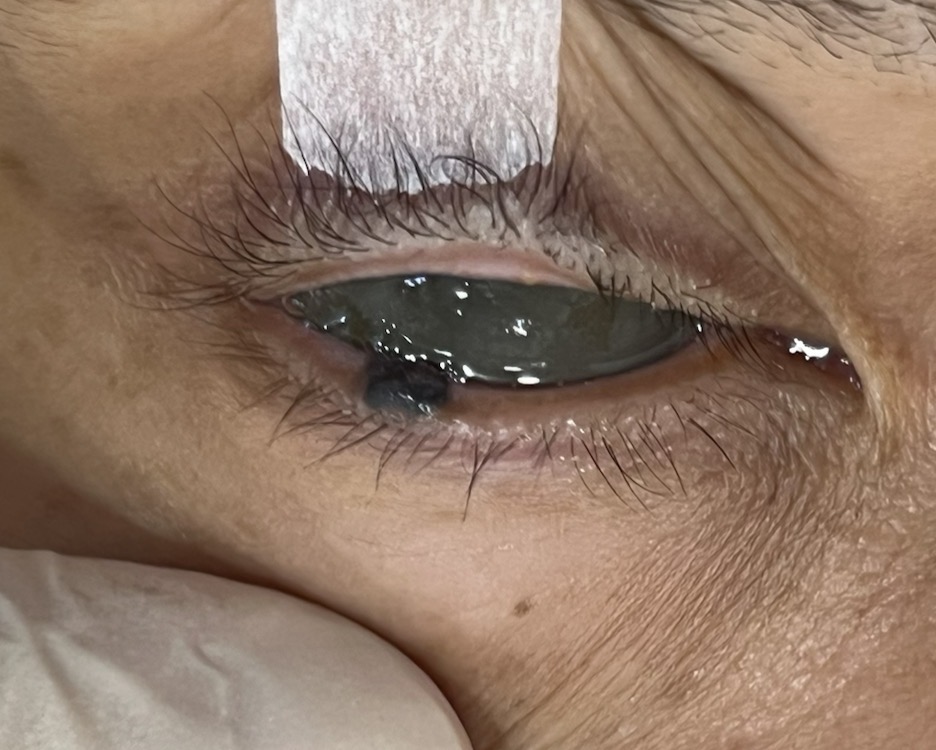
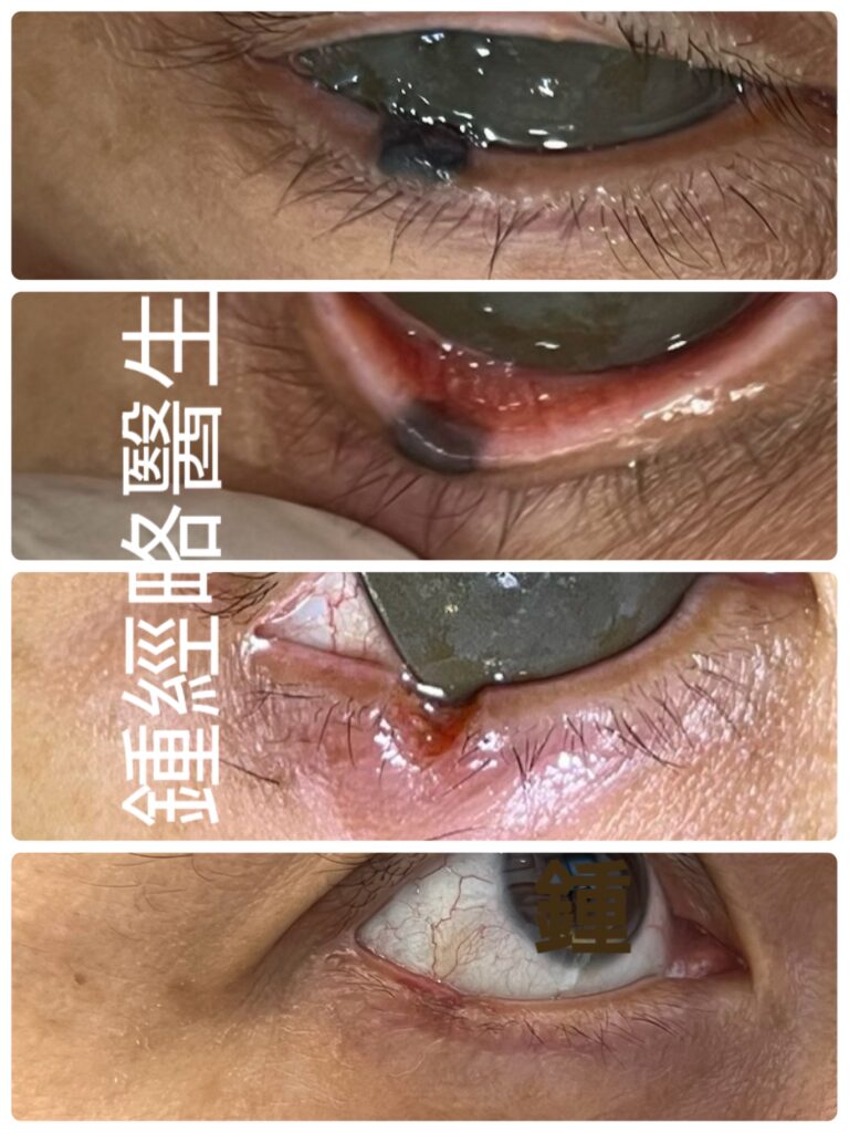
下眼睫邊緣的黑色良性皮膚痣
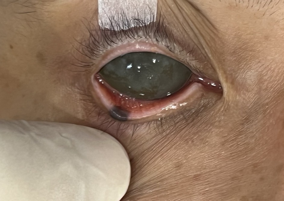
這個色素痣源於下眼皮皮膚,持續跨越眼睫邊緣,接著穿透前葉與後葉,最終深層延伸至眼瞼結膜。
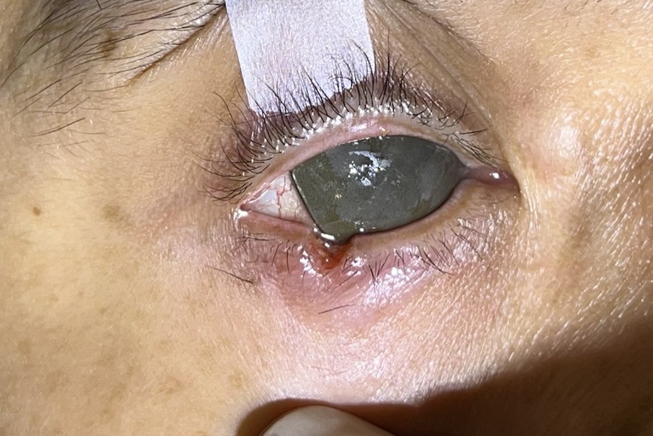
在進行下眼睫邊緣色素痣 CO2 激光脫墨後,下眼瞼出現一個 V 型全層缺損。
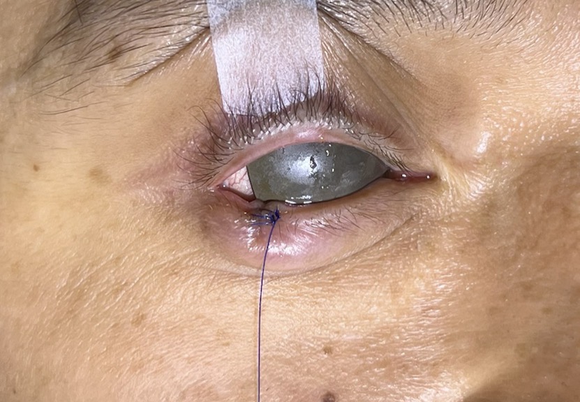
縫合下眼睫邊緣的缺損,再使用 Frost 縫線將下眼瞼向下及向外牽引
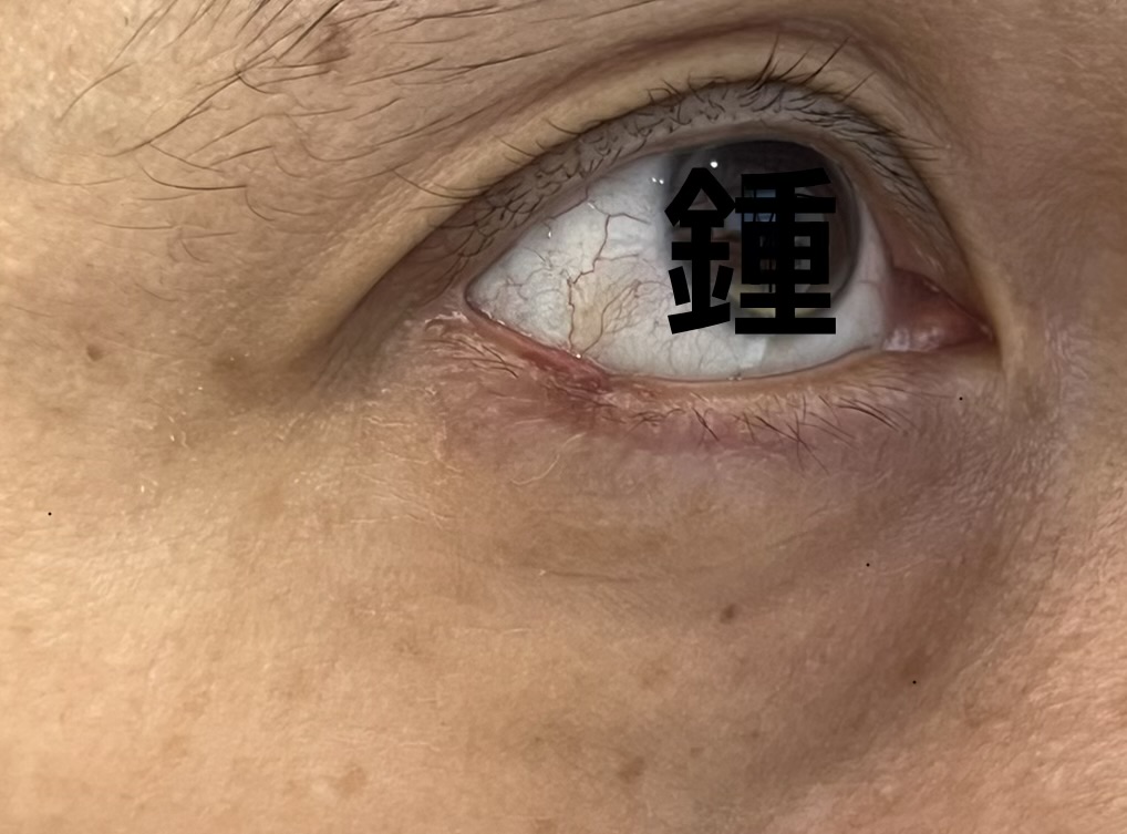
The appearance of the scar on the lower eyelid immediately after suture removal. Scar is expected to fade in the following few months.
拆線後即時顯示下眼瞼疤痕的狀態。預計疤痕將在未來數月內逐漸淡化。
Procedure
- Preparation: The operative field was cleansed with antiseptic solution. Local anesthesia was administered.
- Laser Ablation: A CO₂ laser was used to perform complete ablation of the pigmented lesion. Ablation was carried out layer by layer until normal tissue margins were reached. Ultra-pulse was used to minimise collateral damage. Hemostasis was achieved with the laser.
- Margin Repair:
- Tarsal plate was repaired with absorbable suture.
- A simple interrupted suture was positioned slightly anterior to the gray line to minimize risk of corneal irritation and to ensure precise edge‑to‑edge alignment and prevent margin notching.
- Lower Eyelid Repair: Skin edges were aligned carefully to preserve eyelid contour. Wound was dressed with antibiotic ointment.
Estimated Blood Loss
Minimal.
Complications
None.
Postoperative Wound Care
Ophthalmic Ointments: Apply prescribed antibiotic or lubricating eye ointments regularly. This keeps the suture lines moist and prevents crusting. Additionally, it protects the exposed corneal surface.
Tension and Edema Management: Keep the temporary Frost suture intact for the first 3 to 5 days. Consequently, you will guard against early mechanical pull or spastic inversion while initial fibroplasia takes place. Next, apply cold compresses during the first 48 hours as needed. As a result, you will minimize swelling.
Suture Monitoring and Hygiene: First, gently clean the eyelid margins with sterile saline to remove debris. Meanwhile, avoid rubbing or applying mechanical pressure to the surgical area. Finally, attend scheduled follow-up evaluations to check corneal integrity and ensure timely removal of skin sutures.
Detailed Operative Description of Lower Eyelid Defect Repair with Discussion of Suturing Techniques
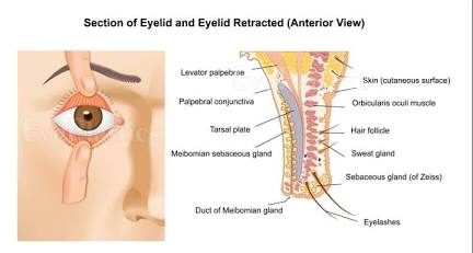
Surgeons often remove eyelid margin lesions using CO2 lasers or traditional surgery. However, this process requires meticulous planning. Ultimately, careful technique preserves both cosmetic appearance and essential eye protection.
Some benign lesions, such as skin moles, extend through both the anterior and posterior lamellae. Consequently, removing them inherently impacts the structural integrity of the eyelid.
Why the Procedure Results in a Full-Thickness Defect
Lesion Extension The lesion’s melanocytes and cellular architecture extend across the entire vertical dimension of the eyelid. Consequently, removing it creates a full-thickness defect. Furthermore, the clinical photograph shows this clearly. The pigmented mole originates on the lower eyelid skin. Next, it spans continuously across the delicate eyelid margin. It then penetrates both the anterior and posterior lamellae. Finally, it reaches deep into the palpebral conjunctiva.
Tissue Eradication and Defect Complete oncologic or aesthetic clearance requires eradicating the entire depth of the lesion. Therefore, surgeons must vaporize or excise tissue from the superficial skin through the tarsal plate and out to the inner mucosal lining. As a result, this process leaves a void that breaches all layers. Ultimately, it creates a full-thickness defect that compromises both the anterior and posterior lamellae.
Risks of Leaving the Defect Untreated or Poorly Repaired
If surgeons fail to close a full-thickness lower eyelid defect, severe complications will occur. Similarly, using a flawed or high-tension repair technique creates major risks. Consequently, these errors expose the globe to both acute and chronic complications:
Corneal Abrasion and Ulceration: Unrepaired structural gaps directly irritate the eye. Similarly, poorly aligned margin knots chronically rub against the corneal epithelium. Consequently, these defects trigger painful abrasions and persistent epithelial defects. Ultimately, they can even cause severe microbial keratitis.
Eyelid Malposition (Ectropion or Entropion): High-tension closures or improper vector alignment often lead to cicatricial contraction. This can cause the eyelid margin to pull outward away from the globe (ectropion). Or it can roll inward so that the eyelashes scratch the surface of the eye (entropion).
Notching and Contour Irregularities: Inadequate multi-layered alignment of the tarsal plate creates a permanent notch or step-off at the eyelid margin. Consequently, this defect disrupts the normal distribution of the tear meniscus. Furthermore, it leads to localized dry spots on the eye surface.
Step-by-Step Full-Thickness Eyelid Margin Repair
Restoring a full-thickness defect demands precise anatomical reconstruction to re-establish structural integrity without abrading the cornea:
1. Tarsal Plate Alignment:
Reapproximate the structural foundation first. Place an interrupted absorbable suture (such as 7-0 Vicryl) through the anterior and mid-depth of the tarsal plate (partial thickness). Keep the suture out of the palpebral conjunctival surface to ensure no rough material contacts the globe.
2. Margin and Gray Line Realignment:
Place a simple interrupted suture (Not the commonly used vertical mattress suture! I prefer using a simple interrupted suture through the gray line. This approach achieves equally good cosmetic and functional outcomes. Furthermore, it enhances blood circulation, promotes faster recovery, and lowers infection risks. Most textbooks emphasize this step. Specifically, placing the first suture through the gray line ensures precise edge-to-edge alignment. Consequently, it prevents a notched margin. Nevertheless, I recommend positioning the first suture slightly anterior to the gray line, farther from the cornea. This adjustment lowers the risk of the suture scratching the corneal surface. Otherwise, the suture can occasionally irritate the eye when it sits too close.
3. Lash Line and Anterior Lamella Closure:
Next, secure a second simple interrupted suture near the base of the eyelashes. Do not use the commonly targeted vertical mattress suture here. Instead, choose a fine non-absorbable material like 6-0 nylon.
4. Knot and Tail Management:
Tie all knots on the anterior skin surface. Next, cut the first/upper most suture tails deliberately long, leaving about 4 to 5 cm. Then, direct these tails downward and outward away from the eye. Consequently, they act as a Frost suture to provide reliable traction support.
5. Traction Support:
Use the uppermost non‑absorbable suture tail as a temporary Frost suture. This provides traction support for the lower eyelid margin by pulling it downward and outward. We secure it with tape to the cheek, thereby neutralizing vertical tension during the early healing phase.
Conclusion
Repair of full‑thickness lower eyelid defects following CO2 laser ablation/excision of margin lesions demands meticulous technique and layered anatomical reconstruction. Because these lesions often traverse both lamellae, their removal inevitably compromises eyelid integrity. Leaving such defects untreated or repairing them poorly risks corneal injury, eyelid malposition, and permanent contour irregularities.
A structured approach restores both function and aesthetics. First, achieve precise tarsal plate alignment. Next, perform careful suturing along the gray line and lash line. Finally, apply thoughtful knot management and traction. Consequently, this process ensures optimal recovery. By prioritizing edge‑to‑edge apposition while minimizing corneal contact, surgeons can achieve stable healing, preserve ocular protection, and maintain cosmetic harmony.
手術紀錄
患者: 53 歲男性
手術名稱: 二氧化碳雷射下眼瞼痣切除並術後縫合
主刀醫師: 鍾經略醫生 MBBS (HK)、MSc Aesthetic Medicine (QMUL)、MSc Clin Derm (Lond)
助理: Joanne
麻醉方式: 2% 利多卡因合併腎上腺素局部浸潤麻醉
主訴
右下眼瞼邊緣有不美觀的皮膚痣。
現病史
患者自述右下眼瞼邊緣有一色素性病灶,存在逾 40 年。病灶緩慢增大,無突變或形態急劇改變。患者否認疼痛、刺激感、分泌物或其他相關症狀。主要因美容考量及個人迷信因素要求切除。
過往病史
- 無重要既往病史
- 無重要家族病史
檢查
- 病灶描述: 均質性黑色皮膚腫塊,大小約 0.4 × 0.2 公分,長軸平行於眼瞼邊緣。
- 隆起程度: 高出皮膚表面約 0.1 公分。
- 範圍: 從下眼瞼皮膚延伸至眼瞼邊緣並深入瞼結膜。
- 臨床表徵: 無紅腫、潰瘍、壓痛或其他繼發性變化。
- 全身檢查: 無異常。
臨床印象
右下眼瞼邊緣良性外觀之色素性病灶,病程長期且無症狀,無可疑惡性特徵。
計畫
- 以手術或雷射切除病灶,目的為美容及個人因素。
- 由於病灶外觀良性,無需進行病理檢驗。
- 提供術後傷口護理指導。
手術過程
準備: 手術區以消毒液清潔,並施行局部麻醉。
雷射消融: 使用二氧化碳雷射對色素病灶進行完整消融。採分層消融方式,直至達到正常組織邊緣。使用超脈衝模式以減少周邊組織損傷。止血由雷射完成。
邊緣修復:
- 以可吸收縫線修復瞼板。
- 在灰線稍前方置入單純間斷縫線,以降低角膜刺激風險,同時確保精確的邊緣對位並避免瞼緣缺刻。
下眼瞼修復: 小心對齊皮膚邊緣以維持眼瞼外形,並以抗生素藥膏覆蓋傷口。
估計失血量
極少。
併發症
無。
術後傷口護理
- 眼科藥膏: 規律塗抹處方抗生素或潤滑眼膏,以保持縫線濕潤,防止結痂,並保護暴露的角膜表面。
- 張力與水腫管理: 術後初期保持臨時 Frost 縫線完整 3 至 5 天,以防止早期機械牽拉或痙攣性內翻,並在纖維母細胞增生初期提供支持。術後 48 小時內可視需要使用冷敷以減少腫脹。
- 縫線監測與衛生: 使用無菌生理鹽水輕柔清潔眼瞼邊緣以去除碎屑。避免揉擦或施加機械壓力於手術部位,並安排隨訪以檢查角膜完整性及確保皮膚縫線及時拆除。
詳細手術描述:下眼瞼缺損修復與縫合技術探討
切除方式
眼瞼邊緣病灶(如二氧化碳雷射切除或外科切除)需謹慎的手術規劃,以同時維持美容外觀與眼球保護功能。當良性病灶(如皮膚痣)延伸至前、後層板時,其切除必然影響眼瞼的結構完整性。
為何手術會造成全層缺損
全層缺損的形成原因在於病灶的黑色素細胞及組織結構橫跨整個眼瞼垂直厚度。臨床照片顯示,起源於下眼瞼皮膚的色素痣連續跨越脆弱的眼瞼邊緣,深入前、後層板並延伸至瞼結膜。完整的腫瘤或美容性清除需徹底去除病灶全層。當消融或切除組織從下眼瞼表皮經過瞼板直至內側黏膜時,所形成的缺口自然破壞所有層次,造成前、後層板的全層缺損。
未修復或修復不良的風險
若未能妥善閉合下眼瞼全層缺損,或採用錯誤的高張力縫合法,將使眼球暴露於急性與慢性併發症:
- 角膜擦傷與潰瘍: 未修復的結構缺口或錯位的縫線結節可能持續摩擦角膜上皮,導致疼痛性擦傷、持續性上皮缺損或細菌性角膜炎。
- 眼瞼位置異常(外翻或內翻): 高張力縫合或不正確的向量排列常導致瘢痕性收縮,使眼瞼邊緣外翻遠離眼球(外翻),或內翻使睫毛刮擦角膜表面(內翻)。
- 缺刻與外形不規則: 瞼板多層縫合不足會造成永久性缺刻或階梯狀變形,破壞淚液分布並導致局部乾燥。
全層下眼瞼修復步驟
修復全層缺損需精確的解剖重建,以恢復結構完整性並避免角膜摩擦:
- 瞼板對位: 先重建結構基礎。於瞼板前部與中層置入可吸收間斷縫線(如 7-0 Vicryl),避免縫線暴露於瞼結膜面以防粗糙材料接觸眼球。
- 邊緣與灰線重建: 在灰線稍前方置入單純間斷縫線(而非常用的垂直褥式縫線)。此法能確保精確的邊緣對位並避免缺刻,同時減少角膜刺激,促進血液循環與癒合。
- 睫毛線與前層板閉合: 在睫毛根部附近以細的不可吸收縫線(如 6-0 nylon)置入第二條單純間斷縫線。
- 結與縫線尾管理: 所有結均打在皮膚表面。最上方的不可吸收縫線尾刻意保留較長(約 4–5 mm),並向下、向外引導,作為 Frost 牽引縫線。
- 牽引支持: 利用最上方縫線尾作為臨時 Frost 縫線,將下眼瞼邊緣向下、向外牽引,並以膠帶固定於臉頰,以中和早期癒合階段的垂直張力。
結論
下眼瞼邊緣病灶經二氧化碳雷射或外科切除後所形成的全層缺損,需精細的分層解剖修復。由於病灶常跨越前、後層板,其切除必然破壞眼瞼完整性。若缺損未修復或修復不良,將導致角膜損傷、眼瞼位置異常及永久性外形缺陷。
採用結構化的修復策略——從瞼板對位開始,逐步完成灰線與睫毛線縫合,並輔以縫線管理與牽引支持——能同時恢復功能與美觀。透過精確的邊緣對位並避免角膜摩擦,外科醫師可確保穩定癒合,維持眼球保護並保有美容效果。