
【注意:本文章及真實病人照片均源自鍾經略醫生的臨床皮膚科碩士及美容醫學碩士課程案例,屬於原創醫學文獻。因此,本院嚴格保留所有版權。同時,我們禁止任何形式的轉載與複製。】
眼瞼邊緣也稱作眼皮邊緣,又稱作睫毛緣,廣東話又叫做眼線位置。位於該處的墨/痣就叫做「淚墨」或「淚痣」。
一顆生長緩慢並帶毛髮的痣 a slow‑growing, hairy mole on the lower eyelid margin.
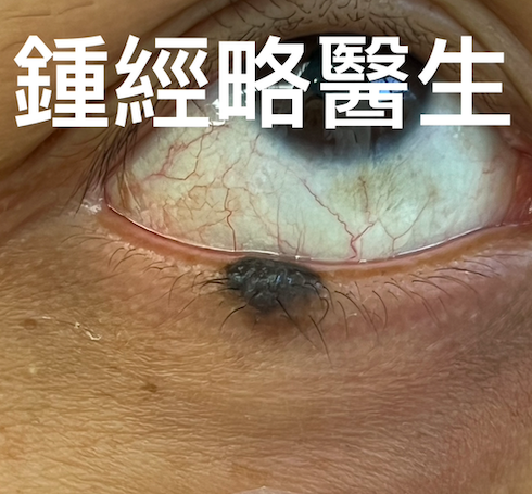
下 眼瞼邊緣有一顆生長緩慢並帶毛髮的痣 a slow‑growing, hairy mole on the lower eyelid margin.
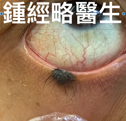
翻轉眼瞼可見黏膜側有色素沉著 the mole extended from the eyelid margin downward into the subcutaneous tissue, reaching the mucosal side of the lower eyelid (the palpebral conjunctiva).
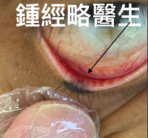
下 眼瞼邊緣脫墨 的激光手術後,下眼瞼邊緣在大概中央位置出現斷裂 Following laser surgery, the lower eyelid margin was disrupted at around the central portion.
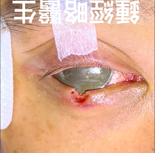
下 眼瞼邊緣脫墨 後怎樣縫合斷開的眼線?以細緻的 6‑0 縫線精確重整眼瞼邊緣。傷口邊緣需極度精準對合,並避免張力,以防止變形
How to suture a broken eyelid margin? The eyelid margin was meticulously realigned using fine 6‑0 sutures. Wound edges were approximated with extreme precision and minimal tension to avoid distortion.
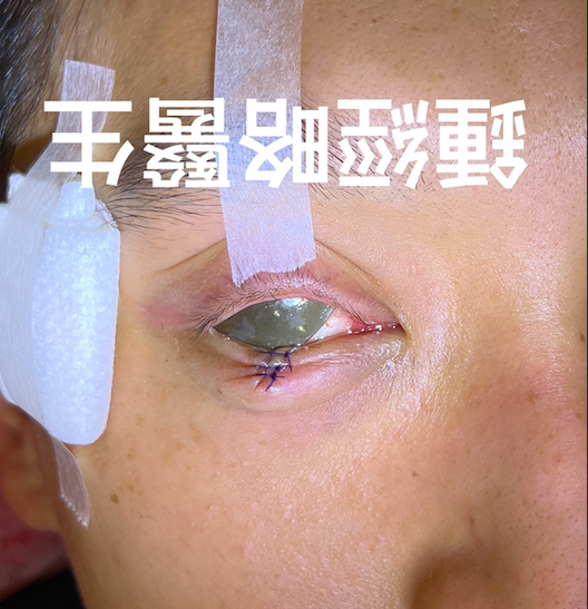
下 眼瞼邊緣脫墨 後傷口縫合後以尼龍縫線輕輕牽拉下眼瞼向外,以避免傷口邊緣摩擦角膜。A nylon suture was placed to gently pull the lower eyelid outward, thereby preventing the wound margin from rubbing against the cornea
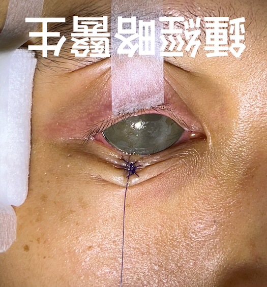
縫線分階段於 2–4 週內拆除 Sutures to be removed in stages over 2–4 weeks.
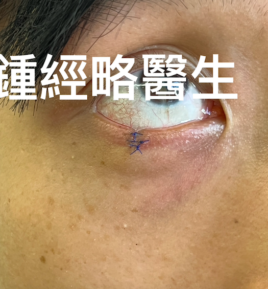
縫線分階段於 2–4 週內拆除 Stitches to be removed in stages over 2–4 weeks.
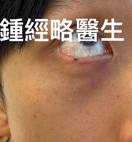
縫線分階段於 2–4 週內拆除 Sutures to be removed in stages over 2–4 weeks.
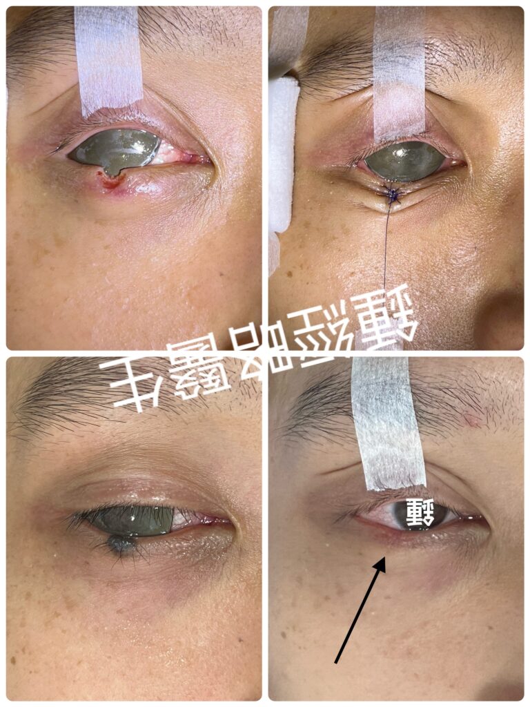
醫療報告:右下眼瞼邊緣脫墨(激光脫淚墨)
術前評估
患者為 35 歲男性,無重要既往病史。下眼瞼邊緣有一顆生長緩慢並帶毛髮的痣。臨床評估顯示為良性病灶;若有不確定性,將進行活檢。術前檢查包括眼瞼解剖及皮膚鬆弛度評估,並規劃切口線。檢查時發現痣自眼瞼邊緣向下延伸至皮下組織,並達到下眼瞼黏膜面。翻轉眼瞼可見黏膜側有色素沉著。若以 CO₂ 激光完全去除痣的「根部」,將不可避免地破壞眼瞼邊緣的連續性,可能導致癒合時間延長、醜陋疤痕,以及外翻或內翻等併發症,影響美觀及眼瞼功能。
因此,我們計劃在 CO₂ 激光消融後,進行精細縫合並準確重整受破壞的下眼瞼邊緣,以避免上述併發症。
安全措施
術中放置潤滑並消毒的內眼盾以保護眼球,具兩大作用:
- 防止 CO₂ 激光造成眼球損傷。
- 防止局部麻醉注射時針頭意外刺傷眼球。已有文獻報告指出,未使用內眼盾時,注射可能導致眼球受傷。
局部麻醉
注射 0.6 ml 含腎上腺素的利多卡因,以減少術中出血。注射前已放置內眼盾。
痣的激光切除(下眼瞼邊緣脫墨)
使用 CO₂ 激光徹底消融痣,包括其基底「根部」,同時盡量保留正常皮膚及眼瞼邊緣。激光手術後,下眼瞼邊緣/眼線在大概中央位置出現斷裂
傷口縫合-怎樣縫合斷開的眼線(斷開的眼瞼邊緣)?
以細緻的 6‑0 縫線精確重整眼瞼邊緣。傷口邊緣需極度精準對合,並避免張力,以防止變形。眼瞼邊緣的準確對合至關重要;若對合不良,將造成眼瞼邊緣「階梯狀」變形,導致美觀缺陷(明顯疤痕、瘢痕疙瘩、皺褶)及功能障礙(眼瞼閉合不全、張開困難、乾眼、雙眼不對稱、眼瞼邊緣摩擦眼球引起不適、角膜損傷或結膜炎)。術後以尼龍縫線輕輕牽拉下眼瞼向外,以避免傷口邊緣摩擦角膜。
眼瞼邊緣脫墨之術後護理
- 切口塗抹抗生素藥膏
- 定期使用潤滑眼藥水
- 密切監測感染及瘢痕情況
- 縫線分階段於 2–4 週內拆除
總結 眼瞼邊緣脫墨獲得良好美容效果的關鍵在於:精確切除、無張力縫合,以及準確重整受破壞的眼瞼邊緣。使用細縫線並配合細心術後護理,可最大程度減少疤痕並維持眼瞼功能。
Medical Report: Laser excision of Benign Mole on Right Lower Eyelid(眼瞼邊緣脫墨)
Preoperative Assessment
A 35‑year‑old male with no significant past medical history presented with a slow‑growing, hairy mole on the lower eyelid margin.
Clinical evaluation confirmed the lesion was benign; if uncertainty arises, biopsy will be performed. Eyelid anatomy and skin laxity were assessed.
On examination, the mole extended from the eyelid margin downward into the subcutaneous tissue, reaching the mucosal side of the lower eyelid. Pigmentation was visible upon eversion of the eyelid. Complete removal of the lesion “root” with CO₂ laser ablation would inevitably disrupt eyelid margin continuity, potentially leading to prolonged healing, unsightly scarring, and complications such as ectropion or entropion, affecting both aesthetics and ocular and eyelid function.
We therefore planned meticulous suturing and precise realignment of the disrupted lower eyelid margin following CO₂ laser ablation, in order to prevent the aforementioned complications.
Safety Measures
A lubricated, sterilized internal eyeshield was placed to protect the globe. This served two purposes:
- Preventing ocular injury from the CO₂ laser.
- Preventing accidental needle trauma during local anesthetic infiltration. Reports exist of inadvertent globe injury when eyelid injections were performed without an internal eyeshield.
Local Anesthesia
0.6 ml of lidocaine with adrenaline was administered to minimize intraoperative bleeding. The eyeshield was in place prior to injection.
Mole Removal
CO₂ laser ablation was used to excise the mole completely, including its base, while preserving as much normal skin and eyelid margin as possible. Following laser surgery, the lower eyelid margin was disrupted at around the central portion.
Wound Closure – how to suture/reconnect a broken eyelid margin?
The eyelid margin was meticulously realigned using fine 6‑0 sutures. Wound edges were approximated with extreme precision and minimal tension to avoid distortion. Accurate apposition of the eyelid margin was critical; poor alignment could result in “stepping” of the margin, leading to cosmetic deformity (visible scar, keloid, wrinkling) and functional impairment (incomplete eyelid closure, lagophthalmos, asymmetry, ocular surface irritation, corneal damage, or conjunctivitis). A nylon suture was placed to gently pull the lower eyelid outward, thereby preventing the wound margin from rubbing against the cornea (Frost suture/traction suture)
Postoperative Care
- Apply antibiotic ointment to the incision.
- Use regular lubricating eyedrops.
- Monitor closely for infection and scarring.
- Sutures to be removed in stages over 2–4 weeks.
Summary
The key to a good cosmetic result on the lower eyelid is precise excision, tension‑free closure, and precise realignment of the disrupted eyelid margin. Using fine sutures and careful postoperative care helps minimize scarring and preserve eyelid function.