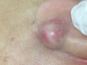
位於左邊耳珠背後的巨大粉瘤。
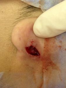
巨大的粉瘤被割除後,留下一個大洞。
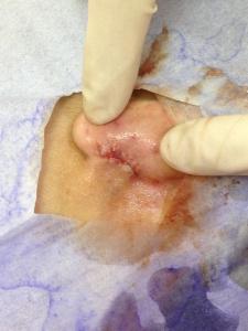
巨大的傷口以6’o (vicryl) 的透明的超幼線縫合,此線會於5個星期自動溶解。
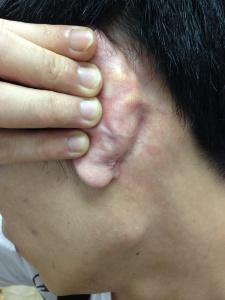
拆線後四個星期,傷口已經完全癒合,傷口並不明顯,也沒有復發的跡象。
An epidermal cyst behind the ear often needs removal because it can enlarge, rupture, become infected, or cause cosmetic and functional problems. Proper excision prevents recurrence and ensures the cyst wall is completely removed.
🔎 Why Removal Is Necessary
- Growth and discomfort: Epidermal cysts are usually benign, but they can slowly enlarge and press on nearby tissue, causing pain or irritation.
- Risk of infection: If the cyst ruptures, keratin material leaks into the dermis, triggering inflammation and sometimes bacterial infection. This can lead to swelling, redness, pus, and scarring.
- Recurrence prevention: Simply draining the cyst is not enough; the cyst wall must be removed. Otherwise, it will refill and recur.
- Cosmetic concerns: Behind the ear, cysts can be visible or palpable, affecting appearance and confidence.
- Rare but important: Although uncommon, epidermal cysts can occasionally be associated with skin cancers, so removal allows histological examination to exclude malignancy.
🩺 Removal Techniques
- Standard surgical excision: The gold standard. A small incision is made, and the entire cyst sac is carefully dissected out intact. This minimizes recurrence.
- Minimal excision technique: A 2–3 mm incision is made, contents expressed, and the cyst wall extracted through the small opening. Less scarring, but technically more challenging.
⚠️ Risks if Not Removed
- Recurrent inflammation: Each rupture or infection makes later removal more difficult due to scar tissue.
- Abscess formation: Untreated infected cysts can form painful abscesses requiring drainage.
- Scarring: Spontaneous rupture often leaves irregular scars, worse than planned excision.
- Psychological impact: Persistent lumps behind the ear can cause anxiety about appearance or health.
✅ Conclusion
Removing an epidermal cyst behind the ear is recommended to prevent infection, recurrence, and scarring, and to allow histological confirmation of its benign nature. Techniques range from standard excision to minimal or laser-assisted methods, with the choice depending on cyst size, inflammation status, and cosmetic considerations. Proper removal ensures safe healing and minimal long-term impact.
耳後表皮囊腫通常需要切除,因為它可能會逐漸增大、破裂、感染,或造成美容及功能上的問題。完整切除能防止復發,並確保囊壁被徹底移除。
🔎 為什麼需要切除
- 增長與不適:表皮囊腫通常是良性的,但它會慢慢增大並壓迫周邊組織,導致疼痛或刺激。
- 感染風險:若囊腫破裂,角蛋白物質滲入真皮,會引起炎症,有時伴隨細菌感染,造成腫脹、紅腫、膿液及疤痕。
- 防止復發:僅僅排出囊腫內容物是不夠的,必須移除囊壁,否則會再次充填並復發。
- 美容困擾:耳後的囊腫可能顯而易見或可觸及,影響外觀與自信。
- 雖然罕見但重要:表皮囊腫偶爾可能與皮膚癌相關,因此切除後進行病理檢查可排除惡性病變。
🩺 切除方法
- 標準外科切除:金標準。做一個小切口,完整剝離囊腫囊壁,降低復發率。
- 微小切口技術:做 2–3 毫米切口,排出內容物,再將囊壁從小開口取出。疤痕較小,但技術上更具挑戰。
⚠️ 不切除的風險
- 反覆炎症:每次破裂或感染都會造成瘢痕組織,使後續切除更困難。
- 膿腫形成:未治療的感染囊腫可能形成疼痛的膿腫,需要引流。
- 疤痕:自發破裂常留下不規則疤痕,比計劃性切除更難看。
- 心理影響:耳後持續存在的腫塊可能引起患者對外觀或健康的焦慮。
✅ 結論
切除耳後表皮囊腫能有效 預防感染、復發與疤痕,並透過病理檢查確認其良性本質。切除方法包括標準外科切除與微小切口技術,選擇依囊腫大小、炎症狀況及美容考量而定。妥善切除可確保安全癒合並減少長期影響。