【注意:本文章及真實病人照片均源自鍾經略醫生的臨床皮膚科碩士及美容醫學碩士課程案例,屬於原創醫學文獻。因此,本院嚴格保留所有版權。同時,我們禁止任何形式的轉載與複製。】
激光脫眉心墨 之案例闡述:
一顆良性痣(墨)位於額頭,位置略高於左眉內側。直徑為0.9公分。從周圍皮膚表面突起約 0.4公分。該病灶為單一、均質的棕色病變,表面平滑,邊界清晰且界線分明。病灶表面可見約 3 至 4 根細毛生長。此痣已存在逾 20 年,除了體積慢慢逐漸變大之外,期間未曾出現顏色、形態或生長模式上的明顯變化。
Case Presentation:
A benign mole is located on the forehead, positioned superior to the medial aspect of the left eyebrow. The solitary, homogenous brown lesion presents with a smooth surface texture and clear, well-defined borders. Approximately 3 to 4 fine hairs emerge from the lesion. The mole has been present for over 20 years, with no reported history of recent changes in color, morphology, or growth pattern.
描述:
位置:位於右眼眉上方,屬於面部顯眼區域。
大小:屬「大墨痣」,估計直徑約為 6–10 毫米,屬中至大型皮膚色素病變。
形狀:邊緣清晰,呈圓形或橢圓形,略呈隆起狀,符合常見的真皮內痣(intradermal nevus)特徵。
顏色:深黑色(墨色),色素分佈均勻,無明顯色差或不規則色斑。
表面質地:表面光滑,無鱗屑或潰瘍,屬良性表徵。
深度:激光手術後形成凹陷傷口,顯示痣根深入真皮層,已被完全清除。
手術方法:激光脫眉心墨:以激光把墨痣徹底連根部一次過切除,根治不復發
術後反應:傷口於三星期內癒合,三個月後疤痕明顯淡化,無明顯色素沉澱或增生。
Description of the mole:
Location: Superior to the eyebrow, on the upper facial region.
Size: Described as a “large mole”, >5 mm in diameter based on clinical terminology.
Shape: Appears well-defined and dome-shaped, typical of benign intradermal nevi.
Color: Deep black, indicating high melanin concentration.
Surface Texture: Smooth and slightly raised, consistent with pigmented lesions.
Depth: The post-laser image reveals a concave wound, suggesting the mole extended into the dermis.
Removal Method: Laser excision, with visible clearance of the mole’s root.
Post-op Observation: Immediate post-surgical photo shows a recessed wound; healing progresses over 3 weeks, with scar fading by 3 months.
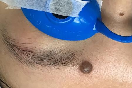
位於眉毛內側(近眉心位置)的一粒深啡色凸出的墨痣
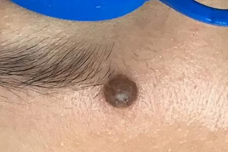
A dark brown raised mole located on the inner side of the eyebrow (near the glabella).
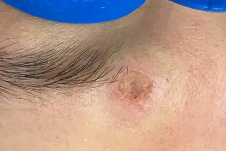
激光脫眉心墨手術後可見皮膚有一凹陷下去的傷口,明顯見到墨的根部已經被完全清除。
After the laser surgery, a sunken wound can be seen on the skin, clearly showing that the root of the mole has been completely removed.
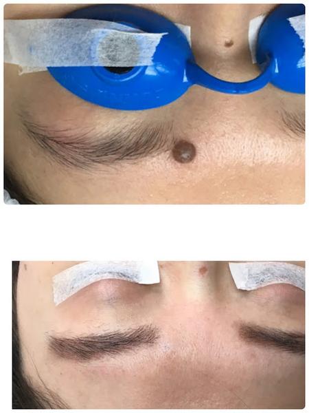
激光脫眉心墨的傷口於3個星期後復原,以下照片於術後3個月後拍攝,可見疤痕已經明顯消退。
The wound healed after three weeks. The following photo was taken three months after the surgery, showing that the scar has already significantly faded
注:在回來覆診時,病人順便把位於鼻樑的一顆較細的墨痣也一併以激光去除。照片中可見鼻樑上有一細小的傷口,那就是脫墨的位置。
Note: During the follow-up visit, the patient also had a smaller mole on the nasal bridge removed with laser. In the photo, a tiny wound can be seen on the nasal bridge, which is the site of the mole removal.
眉心區墨痣切除手術之滑車上動脈與神經的重要性
血管考量
滑車上動脈為前額及眉心區提供血液供應。在進行眉毛上方大痣的手術切除時,若意外損傷該動脈,可能導致大量出血、血腫形成,甚至影響局部循環。因此,醫生必須具備精確的解剖知識,以避免血管損傷。
神經考量
滑車上神經為前額內側及上眼瞼提供感覺。如果在切除過程中損傷該神經,患者可能出現麻木、刺痛或神經性疼痛。此外,神經損傷還會影響面部舒適度與表情,因此在美容性手術中保護神經至關重要。
手術切除的挑戰
深度控制 醫生必須徹底去除痣根部的黑色素細胞,以防復發。然而,深層切除容易造成血管、神經及周圍組織的附帶損傷。
瘢痕風險 眉心區皮膚薄且張力大。結果是,手術縫合往往留下明顯瘢痕,尤其在前額這種表情豐富的部位更為顯眼。
毛囊保護 當痣延伸至眉毛時,切除可能損傷毛囊。因而,患者可能出現眉毛稀疏或不對稱。
激光去痣的優勢
精準與安全 激光消融可逐層靶向黑色素細胞。因此,醫生能更準確地控制深度,並減少對滑車上動脈或神經的附帶損傷。
減少出血 激光在治療過程中可凝固小血管。結果是,出血量遠低於手術切除,通常無需電凝或縫合。
美容效果 激光治療避免大切口,從而減少瘢痕。並且,它能保持周圍皮膚張力與眉毛毛囊,帶來更佳的美學效果。
患者舒適度 激光操作創傷小,通常在局部麻醉下完成。因此,恢復更快,疼痛更輕,兒童或緊張的患者也更容易接受。
結論
滑車上動脈與神經在眉毛上方痣的切除中至關重要。傳統手術切除存在出血、神經損傷、瘢痕及毛囊丟失的風險。相比之下,激光脫眉心墨痣具有更高的精準度、更少的出血、更好的美容效果以及更佳的患者舒適度。因此,在眉心區這種兼顧功能與美觀的部位,激光治療明顯優於傳統手術切除。
The Importance of the Supratrochlear Artery and Nerve in Glabellar Mole Excision
Vascular Considerations
The supratrochlear artery supplies blood to the forehead and glabellar region. During surgical excision of a large mole above the eyebrow, accidental injury to this artery can cause significant bleeding, hematoma formation, or compromise local circulation. Therefore, precise anatomical knowledge is essential to avoid vascular damage.
Neural Considerations
The supratrochlear nerve provides sensation to the medial forehead and upper eyelid. If damaged during excision, patients may experience numbness, tingling, or neuropathic pain. Moreover, nerve injury can affect facial comfort and expression, making preservation critical in cosmetic procedures.
Challenges of Surgical Excision
- Depth Control
- Surgeons must remove all melanocytes at the mole’s root to prevent recurrence.
- However, deep excision risks collateral damage to vessels, nerves, and surrounding tissue.
- Scarring Risk
- The glabellar area has thin skin and high tension.
- As a result, surgical closure often leaves visible scars, especially in expressive regions like the forehead.
- Hair Follicle Preservation
- When the mole extends into the eyebrow, excision can damage hair follicles.
- Consequently, patients may lose eyebrow density or symmetry.
Advantages of Laser Mole Removal
Precision and Safety
Laser ablation allows doctors to target melanocytes layer by layer. Therefore, they can control depth more accurately and reduce the risk of collateral damage to the supratrochlear artery or nerve.
Reduced Bleeding
Lasers coagulate small vessels during treatment. As a result, bleeding is minimal compared to surgical excision, which often requires cauterization or suturing.
Cosmetic Outcomes
Laser treatment minimizes scarring by avoiding large incisions. Moreover, it preserves surrounding skin tension and eyebrow hair follicles, leading to better aesthetic results.
Patient Comfort
Laser procedures are less invasive and often performed under local anesthesia. Consequently, recovery is faster, pain is reduced, and children or anxious patients tolerate the procedure better.
Conclusion
The supratrochlear artery and nerve play a vital role in mole excision above the eyebrow. Surgical removal carries risks of bleeding, nerve injury, scarring, and follicle loss. In contrast, laser mole removal offers superior precision, reduced bleeding, better cosmetic outcomes, and improved patient comfort. Therefore, in the glabellar area—where both function and aesthetics matter most—laser treatment provides clear advantages over traditional surgical excision.