
【注意:本文章及真實病人照片均源自鍾經略醫生的臨床皮膚科碩士及美容醫學碩士課程案例,屬於原創醫學文獻。因此,本院嚴格保留所有版權。同時,我們禁止任何形式的轉載與複製。】
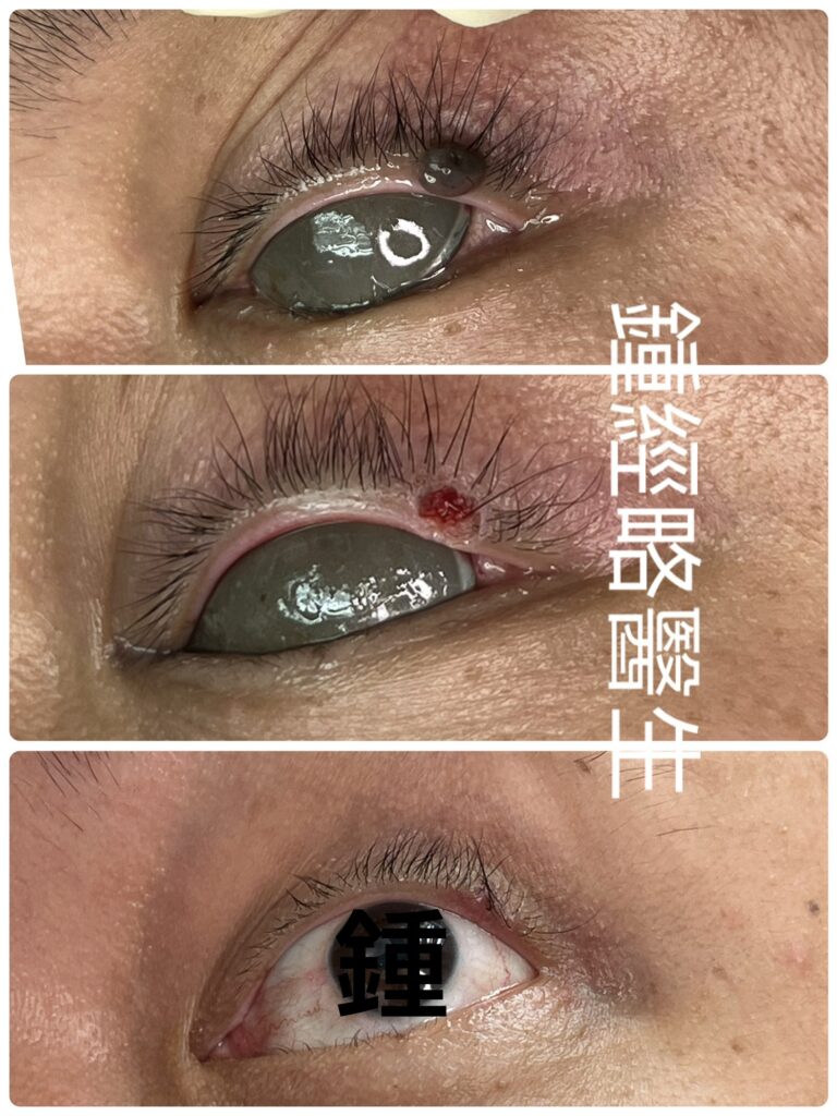
Case Report: CO₂ Laser Removal of a Raised Black Mole on the Left Upper Eyelid Margin
Patient and Lesion Characteristics
A 45‑year‑old woman presented with a raised pigmented mole located on the left upper eyelid margin, just inside the eyelashes. The lesion was:
- Shape: near‑round
- Size: longest diameter 0.3 cm
- Elevation: 0.2 cm above skin level
- Color: uniform black pigmentation
- Surface: smooth
- Margins: well‑defined and clear
The mole had been present for over 15 years and was clinically benign.
Procedure
CO₂ laser ablation was performed to evaporate the entire mole, including its root, to prevent recurrence. After laser treatment, a small crater‑like wound was visible. Notably:
- No bleeding was observed
- No scab or carbonized tissue was present
- The wound floor appeared clean, with no residual root tissue
The depth of ablation was carefully controlled to ensure complete removal while minimizing scarring. Recognition of complete root clearance is based on surgical experience, as the root is subtle and difficult to identify visually.
Surgical Considerations
Determining whether suturing is required after laser mole removal depends on wound depth and tissue involvement. In this case, suturing was deemed unnecessary, as closure might have increased scar formation. The wound was left to heal naturally.
Outcome
At six months post‑procedure, the scar was almost invisible. The cosmetic result was excellent, with no evidence of recurrence.
Discussion
CO₂ laser mole removal offers several advantages:
- Precise vaporization of lesion and root
- Minimal collateral tissue damage
- Reduced risk of recurrence
- Superior cosmetic outcomes compared to traditional excision
However, successful outcomes depend heavily on surgical experience, particularly in judging the depth of ablation and deciding whether suturing is necessary. These skills are difficult to acquire from textbooks or instructional videos and require hands‑on clinical practice.
Important points to consider
This is precisely why a large number of similar cases is essential in training a doctor—only through repeated practice can true skill be developed. Unfortunately, there is currently no reliable way to determine which doctors have undergone such training. In fact, formal training in cosmetic mole removal is minimal in many standard dermatology and plastic surgery curricula.
Therefore, the most practical way to assess a doctor’s expertise is to review the actual cases they have performed. By examining their clinical outcomes, one can judge whether the doctor has genuine experience and proficiency in cosmetic mole removal.
Conclusion
This case demonstrates that CO₂ laser removal of a benign eyelid mole can achieve excellent cosmetic results when performed with careful technique and clinical judgment. Six months after surgery, the scar was nearly invisible, underscoring the effectiveness of laser therapy in delicate facial areas.
病例報告:CO₂ 激光美容治療-左上眼瞼邊緣脫墨(痣)
患者與病灶特徵
患者為 45 歲女性,左上眼瞼邊緣、睫毛內側處有一顆黑色突起痣。病灶特徵如下:
- 形狀:近圓形
- 大小:最長徑 0.3 公分
- 高度:高出皮膚表面約 0.2 公分
- 顏色:均勻黑色
- 表面:光滑
- 邊界:清晰明確
此痣已存在逾 15 年,臨床判斷為良性。
激光手術過程-上眼瞼邊緣脫墨
採用 CO₂ 激光進行汽化切除,將整顆痣連同根部完全去除,以避免復發。術後可見小型凹陷傷口,特點如下:
- 無出血
- 無焦痂或碳化組織殘留
- 傷口底部乾淨,未見殘餘根部
上眼皮邊緣脫墨手術中需精準控制汽化深度,以徹底清除根部並同時減少疤痕形成。判斷根部是否完全去除主要依靠臨床經驗,因其極為細微。
手術考量
是否需要縫合取決於傷口深度及組織受累情況。本例中,縫合並非必要,若強行縫合反而可能增加疤痕形成,因此選擇自然癒合。
術後結果
六個月後回診,疤痕幾乎不可見,美容效果極佳,且未見復發。
討論
CO₂ 激光上眼皮邊緣脫墨具備以下優勢:
- 精準汽化病灶組織,減少周邊損傷
- 能徹底清除根部,降低復發率
- 相較傳統切除手術,美容效果更佳
然而,成功的結果高度依賴臨床經驗,尤其在判斷汽化深度及是否需要縫合方面。這些技巧難以透過教科書或影片學習,必須藉由臨床操作累積。
重點
這正是為什麼醫生在訓練過程中需要大量相似病例,才能真正培養出熟練的技術。可惜的是,目前並沒有任何可靠的方法去辨認哪位醫生接受過這樣的訓練。事實上,在很多的皮膚科或整形外科的常規培訓課程中,幾乎沒有或僅有極少關於美容性去痣(脫墨)的專門訓練。
因此,最實際的方式就是檢視醫生實際完成的病例。透過觀察其臨床成果,才能判斷該醫生是否真正具備美容性去痣(脫墨)的經驗與專業能力。
結論
CO₂ 激光去除良性眼瞼痣(眼皮墨)是一種安全且有效的治療方式。若能精準掌握汽化深度並妥善處理傷口,術後可獲得極佳的美容效果。本例術後六個月,疤痕幾乎不可見,充分顯示激光治療在精細面部區域的優勢。