
目錄 Content
1. 脫墨案例及價錢(按身體部位分類)
👉Case studies & Price (according to body parts)

2. 脫墨最新資訊
👉The Most Update Information on Mole Removal in Hong Kong
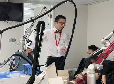
3. 脫墨短片
👉Videos on Mole Removal

4. 脫墨的其他資訊
👉Other information on Mole Removal

🌐 網頁摘要
此網頁重點介紹由鍾經略醫生於其位於香港中環的皮膚科與美容醫學診所所提供的專業脫墨案例。內容包含詳盡的臨床個案研究、手術與激光脫墨技術的參數與說明,以及相關收費參考。
📌 個案研究重點:
每個個案均涵蓋以下特徵分析:
- 痣的位置、形狀、隆起程度、色素分佈及毛髮密度等影像與描述
- 實例包括生長於眼皮、眼線、人中、眉心、頸部、耳朵、嘴唇、鼻尖、鼻樑及鼻側等難做及/或敏感及/或危險部位的痣
- 涵蓋先天性痣及毗鄰動脈(如:滑車上動脈)等高風險位置的個案
- 配合病理檢測結果,以判定痣的性質(良性或惡性)
- 眼部附近進行激光手術時,強調使用眼內保護罩的重要性,研究如何選擇,裝配,潤滑,消毒,儲存眼內保護罩等等
- 使用局部麻醉及無菌技術,所有儀器經高溫滅菌處理,確保在醫療級環境下進行操作,提高病人舒適度與手術安全性
💊鍾經略醫生的專業皮膚科脫墨流程:
當病人前來求診要求去除墨痣時,鍾經略醫生並不會立即進行激光治療,而是採取嚴謹的皮膚科診斷流程,先確認病灶的性質,確定其是否屬於可安全去除的良性病變。只有在完成全面評估後,他才會決定最合適的處理方式——包括觀察、皮膚鏡檢查、活檢、外科切除或激光治療。
- 先問詳細病史
跟其他皮膚科醫生一樣,鍾經略醫生第一步通常會仔細問清楚這粒墨出現了多久、有沒有變大、變色、變凸、痕癢、出血、破損,或者最近有沒有被磨擦到。這一步的重點是分辨它只是普通色素痣,還是有需要高度警惕的皮膚病變 - 精細外觀檢查
之後鍾醫生會直接觀察墨的大小、顏色是否均勻、邊界是否清楚、表面是否對稱,並視乎情況記錄或拍照作日後比較。如果位置在眼皮、手腳、頭皮或其他較難觀察的地方,醫生通常會更仔細評估,因為這些部位的痣不容易自行監察。 - 皮膚鏡檢查分析
很多情況下,鍾醫生會用專業皮膚鏡放大檢查,分析色素分布、血管形態和結構特徵,以判斷是否像良性痣。這是一種非侵入性、通常無痛的檢查方法,是現代皮膚科診斷的重要工具,對決定是否適合進行激光脫墨至關重要。 - 決定是否要化驗
如果外觀不典型、突然變化、位置可疑,或者醫生不能單靠肉眼和皮膚鏡完全判斷,便可能建議切除或取樣化驗。這是因為有些黑色病變不能只靠「美觀」去處理,必須先排除惡性風險。 - 選擇脫墨方法
與其他傳統的/舊派的皮膚科醫生不同,鍾醫生親自執行大部分皮膚外科手術,包括皮膚癌切除、大面積皮膚切除、分層縫合、皮瓣轉移及皮膚移植等,務求為病人提供最佳治療方案,以達致理想效果。
🛠️ 鍾經略醫生提供的專業皮膚科(及皮膚外科)脫墨技術選項:
- 手術切除: 適用於根部深入、毛髮密集或具惡性風險的痣,尤其適合眼皮、眉骨等敏感部位。常見技術包括橢圓形切除、皮瓣移植、皮膚移植及莫氏手術(Mohs surgery)等。
- 二氧化碳激光汽化術(CO₂ Laser Vaporization): 精準且微創,適合處理體積較小、表層淺的痣,減少疤痕並提升美感。
- 分段式手術切除/分階段激光燒灼術: 適用於大型胎記或位置敏感的高風險痣(如靠近眼部),減少一次性創傷,提高癒合效果並降低術後併發症風險。
- 分層傷口縫合:這是一種外科技術,透過逐層縫合各個解剖層來閉合高張力傷口。此方法能提供更強的傷口支撐,促進最佳癒合效果,提升結構穩定性,並改善外觀美感。
- 莫氏手術:這是一種精確的外科技術,在手術過程中運用顯微鏡下的邊緣控制來確保腫瘤完全切除。此技術主要用於治療皮膚癌,特別是基底細胞癌和鱗狀細胞癌,同時亦適用於某些罕見或具侵略性的皮膚癌。莫氏手術特別適合應用於外觀或功能上重要的部位,例如臉部、手部及生殖器等區域。
- 皮瓣修復:常用於修補因切除皮膚病變(如皮膚癌、大型痣、潰瘍或疤痕)所造成的手術缺損,特別適用於無法直接縫合的深層或大面積傷口。透過保留原有的血液供應,皮瓣能有效覆蓋深層或不規則的傷口,並提供更佳的外觀與功能性修復效果。
📖 墨痣的位置(臨床重點示範/皮膚科教學案例)
- 眼眉毛墨痣
- (上/下)眼皮(眼瞼)墨痣
- (上/下)眼瞼邊緣墨痣(眼線墨痣)
- 鼻子墨痣-鼻頭墨痣,鼻翼墨痣,鼻樑墨痣,鼻孔邊緣墨痣
- 額頭墨痣
- 眉心墨痣
- 面頰墨痣
- 鼻唇溝墨痣-笑紋墨痣,虎紋墨痣
- 鬍鬚位置墨痣-上唇白色皮膚部份墨痣(人中墨痣),及下唇白色皮膚部份墨痣
- 嘴唇墨痣(紅色黏膜部分)
- 下巴墨痣-下巴鬍鬚位置墨痣
- 耳朵墨痣-耳珠墨痣,耳殼墨痣,耳孔邊緣墨痣
- 頭皮的墨痣
- 頸墨痣
- 胸口墨痣
- 肚皮墨痣
- 背部墨痣
- 四肢墨痣
- 大動脈附近墨痣
- 大神經附近墨痣
Summary of this webpage:
The webpage centers on the mole removal case studies done by Dr. Chung King Lueh at his dermatology and aesthetic medicine clinic in Central, Hong Kong. It features extensive case studies, clinical descriptions, and fee references for both surgical and laser mole removal. It shows how Dr Chung ensures clearance of a mole while prioritizes minimizing scarring.
Case Study Highlights:
Each case includes mole characteristics like:
- Detailed images and descriptions of mole location, shape, elevation, pigmentation, and hair density.
- Examples include moles on eyelids, philtrum, glabellar region, neck, ears, and nasal area.
- Cases also include congenital moles and those with proximity to arteries (e.g., supratrochlear).
- Histological findings confirming benign/malignant nature
- Clinical safety notes include use of internal eyeshields during laser procedures near the eyes.
- Equipments employed include Local Anaesthesia and sterile technique. Anaesthesia Ensures comfort during both laser and excision procedures. Sterile Technique is employed so that All instruments are autoclaved and procedures are performed in a medical-grade setting.
Dr. Chung King Lueh’s Professional Mole Removal Protocol:
When patients seek mole removal, Dr. Chung King Lueh applies a rigorous dermatological approach rather than proceeding directly to laser treatment. His priority is to establish the precise nature of the lesion and confirm whether it is a benign growth suitable for safe removal. Only after thorough evaluation does he determine the most appropriate management — whether observation, dermoscopy, biopsy, surgical excision, or laser therapy.
- Comprehensive medical history Dr. Chung begins by carefully inquiring about the mole’s duration, any changes in size, color, or elevation, and associated symptoms such as itching, bleeding, or trauma. This step is critical in differentiating a common pigmented nevus from a lesion that may warrant heightened clinical vigilance.
- Detailed visual inspection He then performs a meticulous examination of the mole’s morphology — assessing dimensions, color uniformity, border definition, and surface symmetry. Documentation or clinical photography may be undertaken for longitudinal comparison. Special attention is given to moles located on areas such as the eyelids, scalp, or extremities, where patient self‑monitoring is difficult and professional evaluation is essential.
- Dermoscopic analysis In many cases, Dr. Chung employs dermoscopy to magnify and analyze pigment distribution, vascular structures, and architectural patterns. This non‑invasive, painless technique is a cornerstone of modern dermatology and provides critical information in determining whether a lesion is consistent with benign characteristics and whether laser removal is appropriate.
- Indication for histopathology If the mole exhibits atypical features, undergoes sudden change, arises in a suspicious anatomical site, or cannot be conclusively assessed by clinical and dermoscopic examination, Dr. Chung recommends excision and biopsy for histopathological evaluation. This ensures that potentially malignant lesions are not treated solely for cosmetic purposes, safeguarding patient health before aesthetic considerations are addressed.
- Choosing the best removal method. Unlike many other dermatologists, Dr. Chung personally performs most dermatologic surgical procedures, including skin cancer excisions, large-area skin resections, layered closures, flap transfers, and skin grafts, with the aim of providing patients with the best possible treatment to achieve optimal outcomes.
Techniques Offered by dr chung king lueh:
- Surgical Excision: Recommended for deep, hairy, or suspicious moles; especially effective in sensitive areas like the eyelid and brow. Different excision techniques are discussed eg elliptical excision, skin flap, skin graft, Mohs surgery etc.
- CO₂ Laser Vaporization: Precise and minimally invasive; ideal for smaller and superficial moles.
- Staged Surgical Excision or staged CO2 laser cauterisation: Used for large birthmarks or high-risk moles, particularly near the eyes.
- Layered wound closure: a surgical technique used to close high-tension wounds by suturing each anatomical layer individually. This approach offers stronger wound support and promotes optimal healing, enhances structural integrity, and improves cosmetic outcomes.
- Mohs surgery: a precise surgical technique that uses microscopic margin control during the procedure. It is primarily used to treat skin cancers—especially basal cell carcinoma and squamous cell carcinoma—and is also considered for certain rare or aggressive types of skin cancer. Ideal for cosmetically and functionally critical areas (e.g., face, hands, genitals)
- Skin flaps repair: commonly used to repair surgical defects caused by the removal of skin lesions such as skin cancers, large moles, ulcers, or scars—especially when the wound is too large or deep to close directly. By preserving blood supplies, it covers deep or irregular wounds with better cosmetic and functional outcomes
Various anatomical sites of the mole of concern:
- Eyebrow mole
- Upper/lower eyelid mole
- Upper/lower eyelid margin mole
- Nose mole – nose tip, nose bridge, nose ala
- Forehead mole
- Glabellar area mole
- Cheek mole
- Naso-labial fold mole – smile lines
- Upper/lower cutaneous lip mole – moustache area, beard area
- Upper/lower vermilion lip mole – upper/lower mucosal lip mole
- Chin mole
- Ear mole
- Scalp mole
- Neck mole
- Chest mole
- Abdomen mole
- Back mole
- Limbs mole
- Moles near important/major arteries
- Moles near important/major nerves